. 1999 May;67(5):2110-6.
doi: 10.1128/IAI.67.5.2110-2116.1999.

Role of polymorphonuclear neutrophils in a murine model of Chlamydia psittaci-induced abortion

Affiliations

Role of polymorphonuclear neutrophils in a murine model of Chlamydia psittaci-induced abortion

A J Buendía et al. Infect Immun. 1999 May.

Abstract

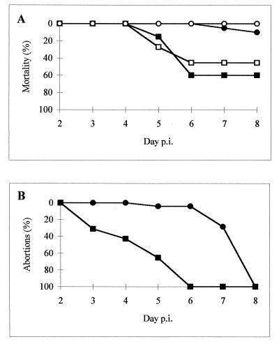
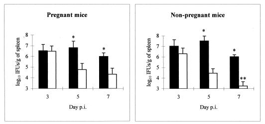
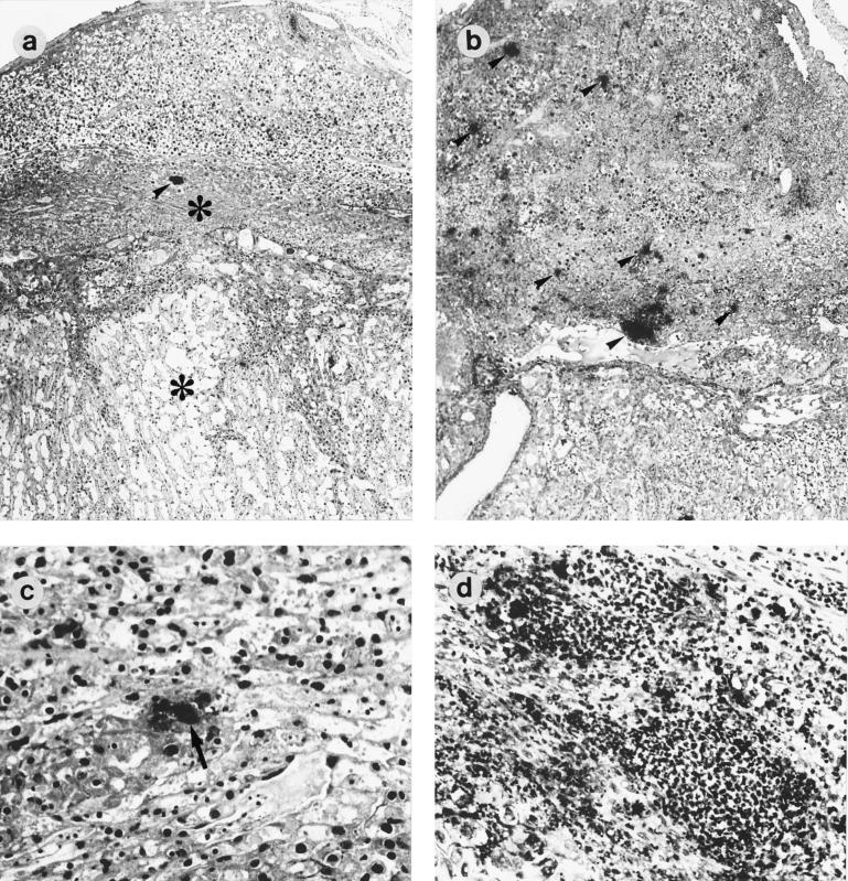
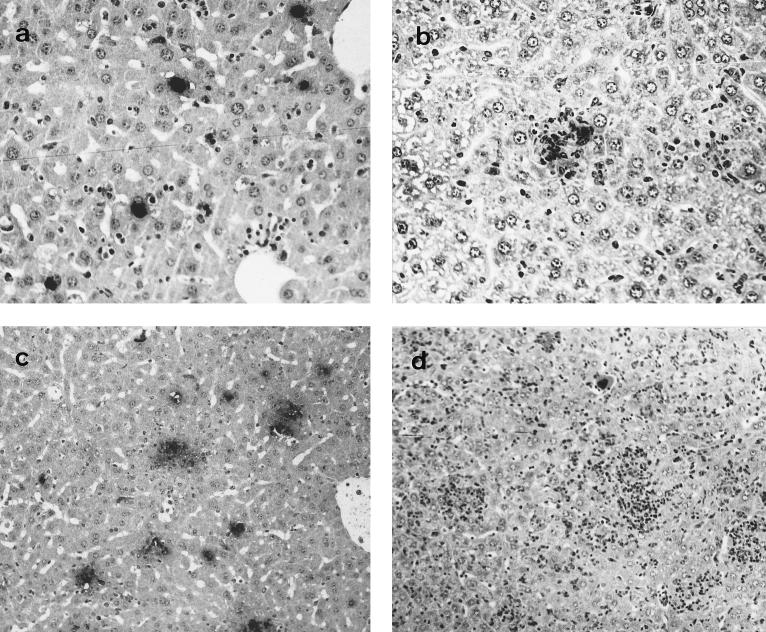
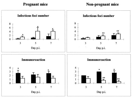
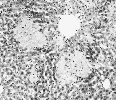
To assess the role of polymorphonuclear neutrophils (PMNs) in Chlamydia psittaci infection in a pregnant mouse model, pregnant and nonpregnant Swiss OF1 mice were depleted of PMNs by treatment with the RB6-8C5 monoclonal antibody before intraperitoneal infection with C. psittaci serotype 1. Nondepleted mice served as infection controls. Depleted mice aborted earlier and had a much higher mortality rate than nondepleted mice. Bacteriological analysis showed that the number of chlamydiae isolated from the spleens of depleted mice at 5 and 7 days postinfection was 100 times greater than that isolated from nondepleted mice. Histopathological analysis of the placentas of depleted mice showed widespread necrosis of the uteroplacental units, with weak immunoreaction to chlamydial antigen, while the placentas of nondepleted mice showed substantial neutrophil infiltration but no large areas of necrosis, with moderate to strong immunoreaction to chlamydial antigen. The livers of depleted mice showed numerous chlamydial inclusions in the hepatocytes, delayed microgranuloma formation, and in the pregnant animals extensive coagulative periportal necrosis. The livers of nondepleted mice displayed multiple small foci of PMNs and mononuclear cells with microgranuloma formation. Among this group of mice, the pregnant animals always had more hepatic damage than nonpregnant animals. Our results suggest that PMNs play an essential role in the response to C. psittaci primary infection, preventing the uncontrolled multiplication of chlamydiae in the liver and spleen.


Figures

FIG. 1
FIG. 1
Administration of antigranulocyte MAbs during C. psittaci infection increases mortality and advances abortions. The animal groups are represented by squares (depleted mice) or circles (nondepleted mice), black for the pregnant mice and white for the nonpregnant mice.
FIG. 2
FIG. 2
Administration of antigranulocyte MAbs increases the growth of C. psittaci in the spleens of infected mice. Shown is the evolution of spleen infection with C. psittaci in neutrophil-depleted (black) and nondepleted (white) groups of pregnant and nonpregnant mice. Quantitative isolation was performed on homogenized tissues from spleens collected at various times p.i. The inclusions were visualized by indirect immunofluorescence. ∗, significant differences (P < 0.05) between depleted and nondepleted mice at the same day p.i.; ∗∗, significant differences (P < 0.05) between pregnant and nonpregnant mice at the same day p.i.
FIG. 3
FIG. 3
Histopathology and immunoreaction in the placentas of mice infected with C. psittaci. Paraffin wax sections immunostained by the ABC method show the distribution of positive immunoreactions (arrowheads and arrows) and areas of necrosis (asterisks) in the placentas of depleted (a and c) and nondepleted (b and d) mice at 5 days p.i. (a) Placenta of a depleted mouse showing only one focus of immunoreaction but extensive areas of necrosis in the decidua basalis and labyrinth; (b) placenta of a nondepleted mouse showing immunoreactive foci spread through the metrial gland and decidua basalis; (c) area of necrosis in the labyrinth of a depleted mouse showing a chlamydial inclusion (arrow) (notice the pyknosis of the nuclei in laberynthine cells); (d) substantial neutrophil infiltration in the decidua basalis of a nondepleted mouse. Magnifications, ×40 (a and b) and ×200 (c and d).
FIG. 4
FIG. 4
Histopathology and immunoreaction in the livers of mice infected with C. psittaci. Paraffin wax sections immunostained by the ABC method show the distribution of positive immunoreactions in the livers of depleted (a and c) and nondepleted (b and d) pregnant mice. (a) Liver of a depleted mouse at 3 days p.i. showing several chlamydial inclusions (notice the lack of leukocytes); (b) liver of a nondepleted mouse at 3 days p.i. showing an infectious focus, formed mainly by neutrophils, with a positive immunoreaction (notice that there are not typical chlamydial inclusions); (c) liver of a depleted mouse at 7 days p.i. showing numerous immunoreactive foci; (d) liver of a nondepleted mouse at 7 days p.i. showing extensive leukocyte infiltration (notice the lack of immunoreactivity in the foci). Magnifications, ×200 (a and b) and ×100 (c and d).
FIG. 5
FIG. 5
Administration of antigranulocyte MAbs during C. psittaci infection decreases the formation of infectious foci but increases the immunoreactivity in the liver. Shown is the evolution of the infection in the liver in the depleted (black) and nondepleted (white) groups of pregnant and nonpregnant mice. Infectious foci were defined as circular lesions formed by leukocytes and were counted in 20 fields (×400 magnification) from a selection of the same lobe of liver from each mouse. The intensity of the immunoreaction is indicated according to the following classification: 0, negative; 1, weak; 2, moderate; 3, strong; and 4, very strong. ∗, significant differences (P < 0.05) between depleted and nondepleted mice at the same day p.i.; ∗∗, significant differences (P < 0.05) between pregnant and nonpregnant mice at the same day p.i.
FIG. 6
FIG. 6
Histopathology in the livers of mice infected with C. psittaci. This paraffin wax section of the liver of a pregnant depleted mouse, stained with hematoxylin-eosin, at 5 days p.i. shows periportal necrosis (light areas). Magnification, ×100.

References

    1. Barteneva N, Theodor I, Peterson E M, de la Maza L M. Role of neutrophils in controlling early stages of a Chlamydia trachomatis infection. Infect Immun. 1996;64:4830–4833. - PMC - PubMed
    1. Baumgärtner W, Bachmann S. Histological and immunocytochemical characterization of Coxiella burnetii-associated lesions in the murine uterus and placenta. Infect Immun. 1992;60:5232–5241. - PMC - PubMed
    1. Buendía A J, Sánchez J, Martínez M C, Cámara P, Navarro J A, Rodolakis A, Salinas J. Kinetics of infection and effects on placental cell populations in a murine model of Chlamydia psittaci-induced abortion. Infect Immun. 1998;66:2128–2134. - PMC - PubMed
    1. Buxton D, Barlow R M, Finlayson J, Anderson I E, Mackellar A. Observations on the pathogenesis of Chlamydia psittaci infection of pregnant sheep. J Comp Pathol. 1990;102:221–237. - PubMed
    1. Cassatella M A. The production of cytokines by polymorphonuclear neutrophils. Immunol Today. 1995;16:21–26. - PubMed

Publication types

MeSH terms

Substances

LinkOut - more resources