. 1999 May;67(5):2184-92.
doi: 10.1128/IAI.67.5.2184-2192.1999.

Cytoskeletal effects induced by pet, the serine protease enterotoxin of enteroaggregative Escherichia coli

Affiliations

Cytoskeletal effects induced by pet, the serine protease enterotoxin of enteroaggregative Escherichia coli

F Navarro-García et al. Infect Immun. 1999 May.

Abstract

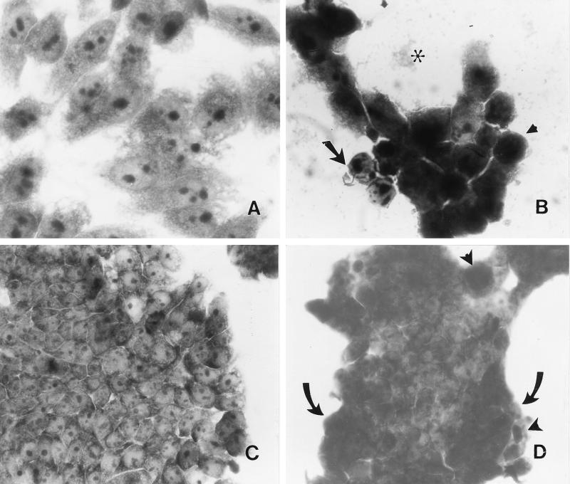
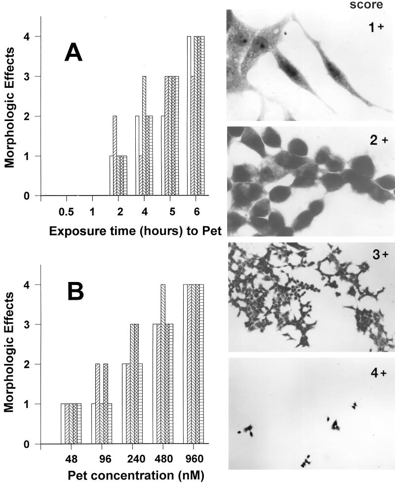
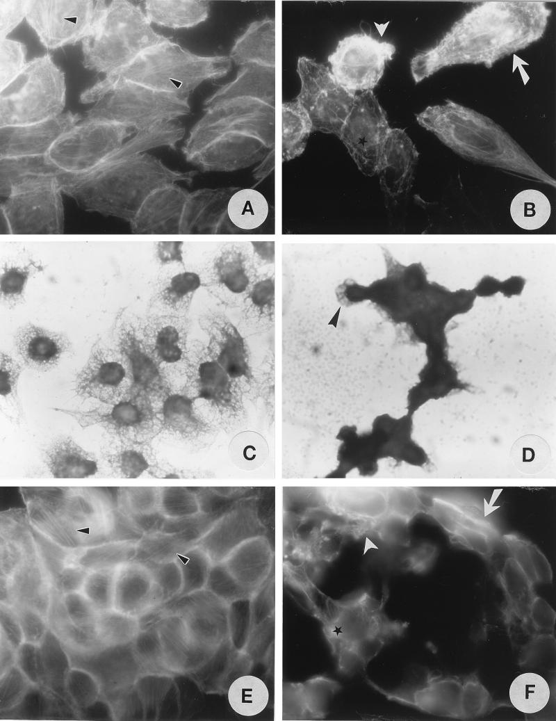
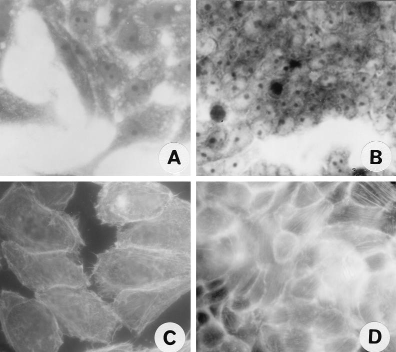
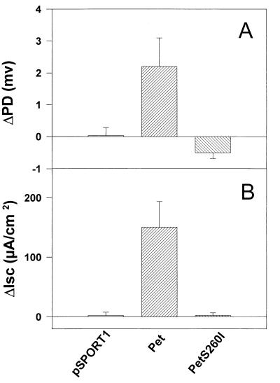
We have previously described enteroaggregative Escherichia coli (EAEC) strains that induce cytotoxic effects on T84 cells, ligated rat ileal loops, and human intestine in culture. Such strains secrete a 104-kDa protein termed Pet (for plasmid-encoded toxin). We have also shown previously that the Pet toxin induces rises in short-circuit current and decreases the electrical resistance in rat jejunum mounted in an Ussing chamber. The nucleotide sequence of the pet gene revealed that Pet is a member of the autotransporter class of secreted proteins. Here we show that a concentrated supernatant of E. coli HB101 harboring the minimal pet clone pCEFN1 induces temperature-, time- and dose-dependent cytopathic effects on HEp-2 cells and HT29 C1 cells in culture. The effects were characterized by release of the cellular focal contacts from the glass substratum, followed by complete rounding of the cells and detachment from the glass. Staining of the Pet-treated cells with Live/Dead viability stain revealed that >90% of rounded cells were viable. Pet-intoxicated HEp-2 and HT29 cells stained with fluorescein-labeled phalloidin revealed contraction of the cytoskeleton and loss of actin stress fibers. However, the effects of Pet were not inhibited by cytoskeleton-altering drugs, including colchicine, taxol, cytochalasin D, and phallicidin. The Pet protein induced proteolysis in zymogram gels, and preincubation with the serine protease inhibitor phenylmethylsulfonyl fluoride resulted in complete abrogation of Pet cytopathic effects. We introduced a mutation in a predicted catalytic serine residue and found that the mutant (Pet S260I) was deficient in protease activity and did not produce cytopathic effects, cytoskeletal damage, or enterotoxic effects in Ussing chambers. These data suggest that Pet is a cytoskeleton-altering toxin and that its protease activity is involved in each of the observed phenotypes.


Figures

FIG. 1
FIG. 1
Effect of Pet protein on epithelial cells. Pet protein was added to cell cultures at a final volume of 500 μl (for HEp-2 cells) or 250 μl (for HT29 cells) per well. Both untreated and treated cells were incubated for 6 h at 37°C in culture medium without antibiotics and serum. (A) HEp-2 cells treated with supernatants from HB101(pSPORT). (B) HEp-2 cells treated with 200 nM Pet protein for 6 h. Release of cellular focal contacts from the glass substratum is indicated with an arrow, rounding of the cells is indicated with an arrowhead, and cell detachment from the glass is indicated with an asterisk. (C) HT29 cells treated with supernatants from HB101(pSPORT). Cells appear normal. (D) HT29 cells treated with 200 nM Pet protein for 6 h. Retraction of the tight cluster is indicated with arrows; rounding and detachment of cells are indicated with arrowheads. Panel B shows remnant cells remaining after near total detachment of cells from the substratum (see text).
FIG. 2
FIG. 2
Quantitation of time- and dose-dependent effects of Pet protein on HEp-2 cells. (A) Time-dependent effects of Pet protein. The effects of Pet protein on the morphology of HEp-2 cells were examined after 0.5, 1, 2, 4, 5, and 6 h of exposure. After this time, monolayers were scored as described in the text. The bars represent individual experiments (n = 4 for each time point). (B) Dose-dependent effects of Pet protein. HEp-2 cells were exposed to different concentrations of Pet protein (48 to 960 nM). The bars represent individual experiments (n = 4 for each concentration point). The inserts show the score of the morphologic changes of HEp-2 cells produced by Pet protein described in Materials and Methods.
FIG. 3
FIG. 3
Effects of Pet protein on the epithelial cell cytoskeleton as detected by FAS (A, B, E, and F) and Coomassie blue staining (C and D). (A and C) Untreated HEp-2 cells. (B and D) HEp-2 cells treated with 200 nM Pet protein for 6 h. (E) Untreated HT29 cells. (F) HT29 cells treated with 200 nM Pet protein for 6 h. Linear F-actin stress fibers are indicated by small arrowheads, contraction of the cytoskeleton by is indicated arrows, loss of actin stress fibers by is indicated asterisks, and formation of surface blebs by is indicated large arrowheads.
FIG. 4
FIG. 4
Proteolytic activity of Pet protein. (A) Sodium dodecyl sulfate-polyacrylamide gel electrophoresis of the 104-kDa secreted Pet protein obtained from the supernatant of the minimal clone HB101(pCEFN1). An L-broth culture was grown overnight at 37°C, and then the supernatant was concentrated and fractionated with a 100-kDa-retention filter. Sizes of marker proteins are indicated in kilodaltons on the right. (B) Proteolytic activity detected by electrophoresing Pet protein alone or mixed with PMSF inhibitor in a precast gelatin zymogram gel. Protease activities are identified as clear bands against a dark blue background after staining with Coomassie blue.
FIG. 5
FIG. 5
Effects of Pet S260I mutant on epithelial cells visualized by Coomassie blue (A and B) or FAS (C and D). (A) HEp-2 cells treated with 96 nM S260I protein for 5 h. (B) HT29 cells treated with 96 nM S260I protein for 5 h. (C) HEp-2 cells treated with 96 nM S260I protein for 5 h. (D) HT29 cells treated with 96 nM S260I protein for 5 h.
FIG. 6
FIG. 6
Effect of the S260I mutation on Pet enterotoxicity. Supernatants from overnight cultures were size fractionated (>100 kDa), and 25 μg of protein was added to each Ussing chamber, mounted with full-thickness rat jejunal tissue. Supernatants from HB101(pCEFN1) and HB101(pSPORT1) were used as a positive and negative controls, respectively. Data points represent the means of at least four experiments; error bars represent standard errors of the mean. The supernatant of pCEFN1 (Pet protein) generates significant rises in PD and Isc compared with the S260I mutant (P < 0.05 by Student’s t test).

References

    1. Benjelloun-Touimi Z, Tahar M S, Montecucco C, Sansonetti P J, Parsot C. SepA, the 110 kDa protein secreted by Shigella flexneri: two-domain structure and proteolytic activity. Microbiology. 1995;144:1815–1822. - PubMed
    1. Bhan M K, Raj P, Levine M M, Kaper J B, Bhandari N, Srivastava R, Kumar R, Sazawal S. Enteroaggregative Escherichia coli associated with persistent diarrhea in a cohort of rural children in India. J Infect Dis. 1989;159:1061–1064. - PubMed
    1. Brunder W, Schmidt H, Karch H. EspP, a novel extracellular serine protease of enterohaemorrhagic Escherichia coli O157:H7 cleaves human coagulation factor V. Mol Microbiol. 1997;24:767–78. - PubMed
    1. Cravioto A, Tello A, Navarro A, Ruiz J, Villafan H, Uribe F, Eslava C. Association of Escherichia coli HEp-2 adherence patterns with type and duration of diarrhoea. Lancet. 1991;337:262–264. - PubMed
    1. de Bernard M, Papini E, de Filippis V, Gottardi E, Telford J, Manetti R, Fontana A, Rappuoli R, Montecucco C. Low pH activates the vacuolating toxin of Helicobacter pylori, which becomes acid and pepsin resistant. J Biol Chem. 1995;270:23937–23940. - PubMed

Publication types

MeSH terms

LinkOut - more resources