. 1999 May;67(5):2349-56.
doi: 10.1128/IAI.67.5.2349-2356.1999.

Gamma interferon production is critical for protective immunity to infection with blood-stage Plasmodium berghei XAT but neither NO production nor NK cell activation is critical

Affiliations

Gamma interferon production is critical for protective immunity to infection with blood-stage Plasmodium berghei XAT but neither NO production nor NK cell activation is critical

T Yoneto et al. Infect Immun. 1999 May.

Abstract

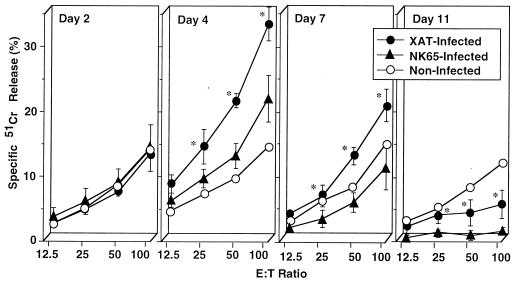
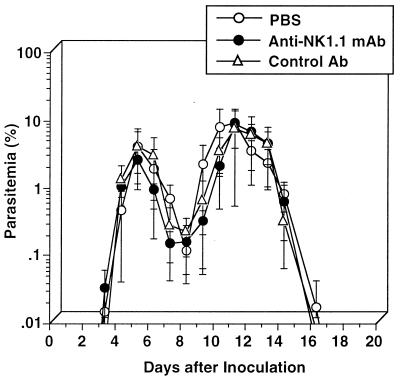
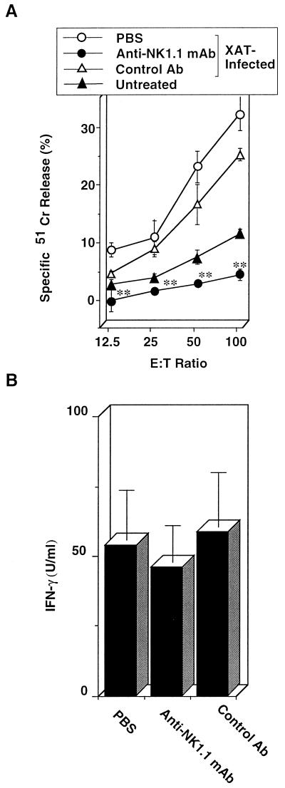
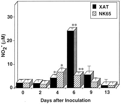
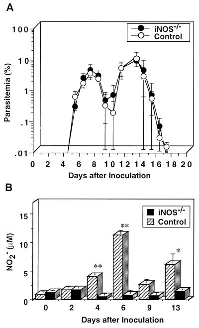
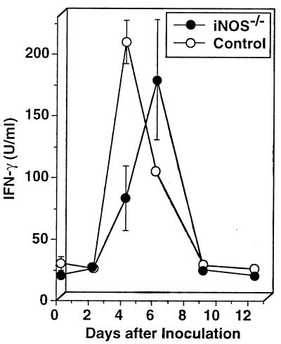
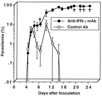
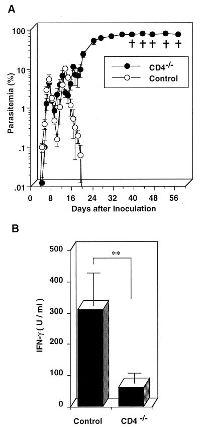
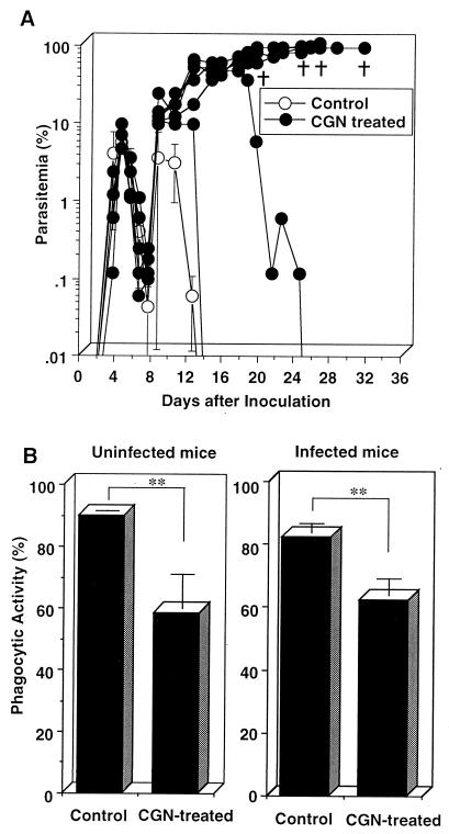
We have examined the roles of gamma interferon (IFN-gamma), nitric oxide (NO), and natural killer (NK) cells in the host resistance to infection with the blood-stage malarial parasite Plasmodium berghei XAT, an irradiation-induced attenuated variant of the lethal strain P. berghei NK65. Although the infection with P. berghei XAT enhanced NK cell lytic activity of splenocytes, depletion of NK1.1(+) cells caused by the treatment of mice with anti-NK1.1 antibody affected neither parasitemia nor IFN-gamma production by their splenocytes. The P. berghei XAT infection induced a large amount of NO production by splenocytes during the first peak of parasitemia, while P. berghei NK65 infection induced a small amount. Unexpectedly, however, mice deficient in inducible nitric oxide synthase (iNOS-/-) cleared P. berghei XAT after two peaks of parasitemia were observed, as occurred for wild-type control mice. Although the infected iNOS-/- mouse splenocytes did not produce a detectable level of NO, they produced an amount of IFN-gamma comparable to that produced by wild-type control mouse splenocytes, and treatment of these mice with neutralizing anti-IFN-gamma antibody led to the progression of parasitemia and fatal outcome. CD4(-/-) mice infected with P. berghei XAT could not clear the parasite, and all these mice died with apparently reduced IFN-gamma production. Furthermore, treatment with carrageenan increased the susceptibility of mice to P. berghei XAT infection. These results suggest that neither NO production nor NK cell activation is critical for the resistance to P. berghei XAT infection and that IFN-gamma plays an important role in the elimination of malarial parasites, possibly by the enhancement of phagocytic activity of macrophages.


Figures

FIG. 1
FIG. 1
Increased NK cell lytic activity in splenocytes caused by the infection with blood-stage P. berghei XAT but not with P. berghei NK65. After i.v. inoculation of normal mice with 104 PRBC, splenocytes were obtained at various time intervals and assayed for NK cell lytic activity by using YAC-1 cells as a target. The percentages of spontaneous 51Cr release in assays of splenocytes obtained on days 2, 4, 7, and 11 were 5.7, 6.1, 10.7, and 5.7% of the maximum release, respectively. Data are means ± SD for three mice. ∗, P < 0.05, compared with data for NK65-infected mice. These results were confirmed to be reproducible by performing a repeat experiment. E:T, effector-to-target-cell.
FIG. 2
FIG. 2
No effect of NK cell depletion on the resistance to infection with blood-stage P. berghei XAT. NK1.1+ cells were depleted by treatment with anti-NK1.1 MAb at 0.3 mg/injection/mouse once daily for 3 consecutive days before the day of inoculation i.v. of 104 PRBC and then every other day for 20 days. Normal rat IgG was used as a control antibody. Parasitemia was assessed by the microscopic examination of Giemsa-stained smears of tail blood. Data are means ± SD for five mice. These results were confirmed to be reproducible by performing a repeat experiment.
FIG. 3
FIG. 3
NK cell lytic activity was reduced in mice treated with anti-NK1.1 without impairment of IFN-γ production. Mice were injected i.p. at 0.3 mg/injection/mouse with anti-NK1.1 once daily for 3 consecutive days before the day of the 1 × 10⁴ PRBC i.v. inoculation and then every other day for 20 days. (A) Spleens were obtained 4 days after the inoculation of parasites and assayed for NK cell lytic activity by using YAC-1 cells as a target. E:T, effector-to-target-cell. The percent spontaneous 51Cr release in the assay was 8.7% of the maximum release. Data are means ± SD for three mice. (B) Splenocytes obtained 4 days after the parasite inoculation were cultured without addition of parasite antigen for 48 h, and the culture supernatants were assayed for IFN-γ by using an ELISA. Data are means ± SD for three mice. ∗∗, P < 0.01, compared with the data in PBS- or control antibody-treated mice. These results were confirmed to be reproducible by performing a repeat experiment.
FIG. 4
FIG. 4
Splenocytes obtained from mice infected with blood-stage P. berghei XAT produced more NO than those obtained from mice infected with P. berghei NK65. After i.v. inoculation of 104 PRBC, splenocytes were obtained at various intervals and cultured in vitro without addition of parasite antigen for 72 h. The culture supernatants were assayed for NO2. Data are means ± SD for three mice. ∗, P < 0.05, and ∗∗, P < 0.01, compared with the data for splenocytes obtained on day 0. Similar experiments were performed three times, and essentially the same results were obtained.
FIG. 5
FIG. 5
Comparable susceptibilities of iNOS−/− mice and wild-type control mice to the infection with blood-stage P. berghei XAT. (A) After iNOS−/− mice or wild-type control mice were inoculated i.v. with 104 PRBC, parasitemia was assessed by the microscopic examination of Giemsa-stained smears of tail blood. Data are means ± SD for five mice. (B) After iNOS−/− mice or wild-type control mice were inoculated i.v. with 104 PRBC, splenocytes were obtained at various intervals and cultured in vitro without addition of parasite antigen for 72 h. The culture supernatants were assayed for NO2. Data are means ± SD for three mice. ∗, P < 0.05, and ∗∗, P < 0.01, compared with the data for splenocytes obtained on day 0. These experiments were performed three times, and similar results were obtained.
FIG. 6
FIG. 6
Comparable production of IFN-γ in splenocytes from iNOS−/− mice and in those from wild-type control mice caused by the infection with blood-stage P. berghei XAT. After iNOS−/− mice or wild-type control mice were inoculated i.v. with 104 PRBC, splenocytes were obtained at various intervals and cultured in vitro without addition of parasite antigen for 48 h. The culture supernatants were assayed for IFN-γ by using an ELISA. Data are means ± SD for three mice. Similar results were obtained in two successive experiments.
FIG. 7
FIG. 7
Important role for IFN-γ in the host resistance of iNOS−/− mice against the infection with blood-stage P. berghei XAT. After iNOS−/− mice were inoculated i.v. with 104 PRBC, endogenously produced IFN-γ was neutralized by treatment at 0.2 mg/mouse with anti-IFN-γ once daily for 4 consecutive days starting from the day of the inoculation and then twice a week for 3 weeks. Parasitemia was assessed by the microscopic examination of Giemsa-stained smears of tail blood. Normal rat IgG was used as a control antibody. Data are means ± SD for five mice. †, days on which individual mice died. We obtained essentially the same results in three successive experiments.
FIG. 8
FIG. 8
Increased susceptibility to blood-stage P. berghei XAT infection of CD4−/− mice with reduced IFN-γ production. (A) After CD4−/− mice or wild-type control mice were inoculated i.v. with 104 PRBC, parasitemia was assessed by the microscopic examination of Giemsa-stained smears of tail blood. Data are means ± SD for five mice. †, days on which individual mice died. (B) Splenocytes were obtained on day 4 after the parasite inoculation and cultured in vitro without addition of parasite antigen for 48 h. The culture supernatants were assayed for IFN-γ by using an ELISA. Data are means ± SD for three mice. ∗∗, P < 0.01. The results presented in both panels A and B were confirmed to be reproducible in two successive experiments.
FIG. 9
FIG. 9
Increased susceptibility of mice treated with CGN to blood-stage P. berghei XAT infection with reduced phagocytic activity of spleen macrophages. (A) Mice treated with CGN as described in Materials and Methods were inoculated i.v. with 104 PRBC, and parasitemia was assessed by the microscopic examination of Giemsa-stained smears of tail blood. †, days on which individual mice died. (B) Adherent spleen cells obtained from CGN-treated uninfected mice and also from mice infected for 14 days were incubated with FITC-conjugated beads for 2 h. Phagocytic activity was assayed by FACScan analysis with gating on a Mac-1high population. Data are means ± SD for eight mice. ∗∗, P < 0.01. These results were confirmed to be reproducible in two successive experiments.

References

    1. Bancroft G J. The role of natural killer cells in innate resistance to infection. Curr Opin Immunol. 1993;5:503–510. - PubMed
    1. Brown K M, Kreier J P. Effect of macrophage activation on phagocyte-plasmodium interaction. Infect Immun. 1986;51:744–749. - PMC - PubMed
    1. Buffinton G D, Hunt N H, Cowden W B, Clark I A. Detection of short-chain carbonyl products of lipid peroxidation from malaria-parasite (Plasmodium vinckei)-infected red blood cells exposed to oxidative stress. Biochem J. 1988;249:63–68. - PMC - PubMed
    1. Cherwinski H M, Schumacher J H, Brown K D, Mosmann T R. Two types of mouse helper T cell clone. III. Further differences in lymphokine synthesis between Th1 and Th2 clones revealed by RNA hybridization, functionally monospecific bioassays, and monoclonal antibodies. J Exp Med. 1987;166:1229–1244. - PMC - PubMed
    1. Clark I A, Butcher G A, Buffinton G D, Hunt N H, Cowden W B. Toxicity of certain products of lipid peroxidation to the human malaria parasite Plasmodium falciparum. Biochem Pharmacol. 1987;36:543–546. - PubMed

Publication types

MeSH terms