

Enterohemorrhagic Escherichia coli O157:H7 produces Tir, which is translocated to the host cell membrane but is not tyrosine phosphorylated
- PMID: 10225900
- PMCID: PMC115983
- DOI: 10.1128/IAI.67.5.2389-2398.1999
Enterohemorrhagic Escherichia coli O157:H7 produces Tir, which is translocated to the host cell membrane but is not tyrosine phosphorylated
Abstract
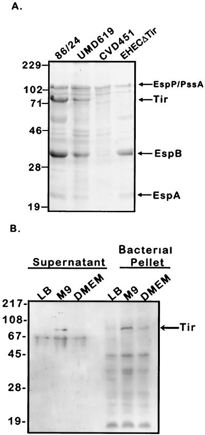
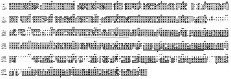
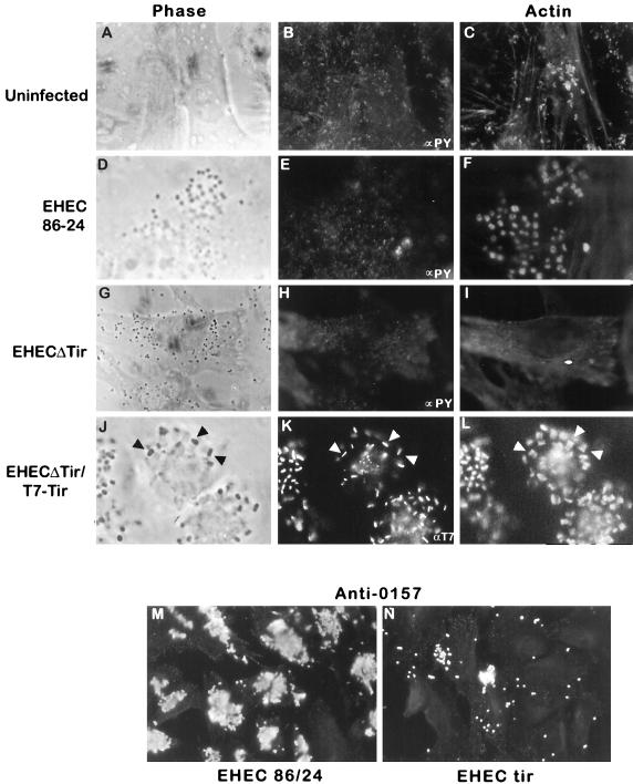
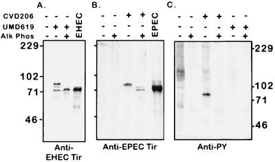
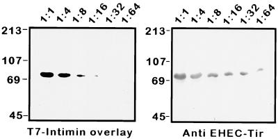
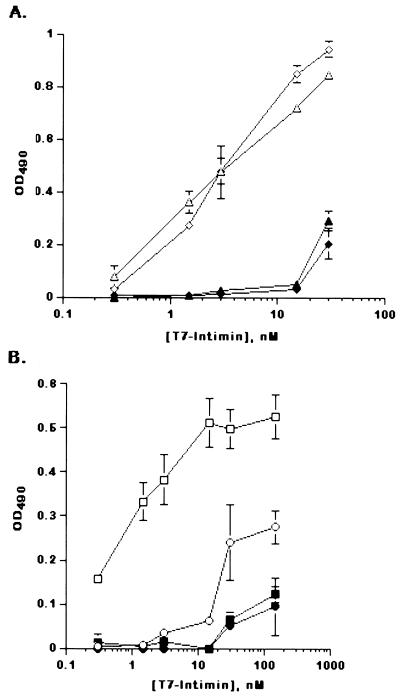
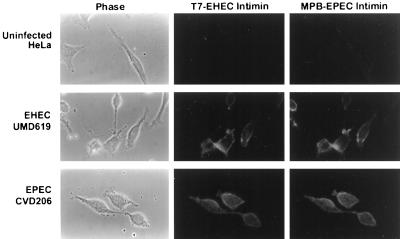
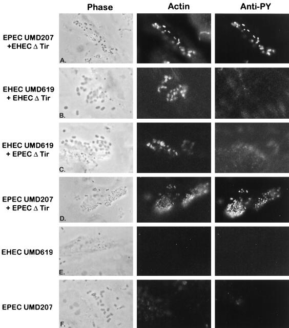
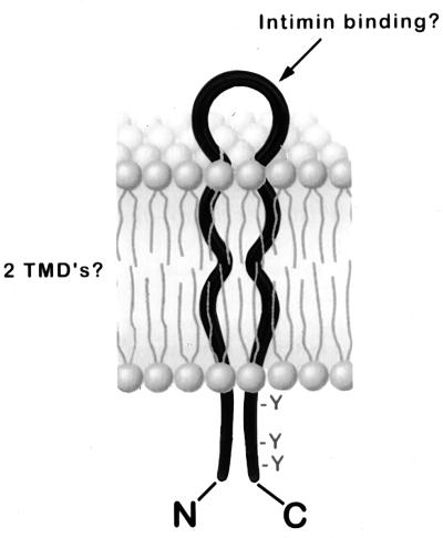
Intimate attachment to the host cell leading to the formation of attaching and effacing (A/E) lesions is an essential feature of enterohemorrhagic Escherichia coli (EHEC) O157:H7 pathogenesis. In a related pathogen, enteropathogenic E. coli (EPEC), this activity is dependent upon translocation of the intimin receptor, Tir, which becomes tyrosine phosphorylated within the host cell membrane. In contrast, the accumulation of tyrosine-phosphorylated proteins beneath adherent EHEC bacteria does not occur, leading to questions about whether EHEC uses a Tir-based mechanism for adherence and A/E lesion formation. In this report, we demonstrate that EHEC produces a functional Tir that is inserted into host cell membranes, where it serves as an intimin receptor. However, unlike in EPEC, in EHEC Tir is not tyrosine phosphorylated yet plays a key role in both bacterial adherence to epithelial cells and pedestal formation. EHEC, but not EPEC, was unable to synthesize Tir in Luria-Bertani medium but was able to secrete Tir into M9 medium, suggesting that Tir synthesis and secretion may be regulated differently in these two pathogens. EHEC Tir and EPEC Tir both bind intimin and focus cytoskeletal rearrangements, indicating that tyrosine phosphorylation is not needed for pedestal formation. EHEC and EPEC intimins are functionally interchangeable, but EHEC Tir shows a much greater affinity for EHEC intimin than for EPEC intimin. These findings highlight some of the differences and similarities between EHEC and EPEC virulence mechanisms, which can be exploited to further define the molecular basis of pedestal formation.
Figures
Similar articles
-
Phosphorylation of tyrosine 474 of the enteropathogenic Escherichia coli (EPEC) Tir receptor molecule is essential for actin nucleating activity and is preceded by additional host modifications.Mol Microbiol. 1999 Feb;31(4):1229-41. doi: 10.1046/j.1365-2958.1999.01265.x. Mol Microbiol. 1999. PMID: 10096089
-
Enterohaemorrhagic and enteropathogenic Escherichia coli use a different Tir-based mechanism for pedestal formation.Mol Microbiol. 2001 Sep;41(6):1445-58. doi: 10.1046/j.1365-2958.2001.02617.x. Mol Microbiol. 2001. PMID: 11580847
-
Enteropathogenic E. coli Tir binds Nck to initiate actin pedestal formation in host cells.Nat Cell Biol. 2001 Sep;3(9):856-9. doi: 10.1038/ncb0901-856. Nat Cell Biol. 2001. PMID: 11533668
-
Mechanism of action of EPEC type III effector molecules.Int J Med Microbiol. 2002 Feb;291(6-7):469-77. doi: 10.1078/1438-4221-00155. Int J Med Microbiol. 2002. PMID: 11890546 Review.
-
Tails of two Tirs: actin pedestal formation by enteropathogenic E. coli and enterohemorrhagic E. coli O157:H7.Curr Opin Microbiol. 2003 Feb;6(1):82-90. doi: 10.1016/s1369-5274(03)00005-5. Curr Opin Microbiol. 2003. PMID: 12615225 Review.
Cited by
-
Actin cytoskeleton manipulation by effector proteins secreted by diarrheagenic Escherichia coli pathotypes.Biomed Res Int. 2013;2013:374395. doi: 10.1155/2013/374395. Epub 2012 Dec 30. Biomed Res Int. 2013. PMID: 23509714 Free PMC article. Review.
-
CEACAM6 acts as a receptor for adherent-invasive E. coli, supporting ileal mucosa colonization in Crohn disease.J Clin Invest. 2007 Jun;117(6):1566-74. doi: 10.1172/JCI30504. Epub 2007 May 24. J Clin Invest. 2007. PMID: 17525800 Free PMC article.
-
The StcE protease contributes to intimate adherence of enterohemorrhagic Escherichia coli O157:H7 to host cells.Infect Immun. 2005 Mar;73(3):1295-303. doi: 10.1128/IAI.73.3.1295-1303.2005. Infect Immun. 2005. PMID: 15731026 Free PMC article.
-
Characterization of the binding surface of the translocated intimin receptor, an essential protein for EPEC and EHEC cell adhesion.Protein Sci. 2007 Dec;16(12):2677-83. doi: 10.1110/ps.073128607. Protein Sci. 2007. PMID: 18029421 Free PMC article.
-
Heterogeneous surface expression of EspA translocon filaments by Escherichia coli O157:H7 is controlled at the posttranscriptional level.Infect Immun. 2003 Oct;71(10):5900-9. doi: 10.1128/IAI.71.10.5900-5909.2003. Infect Immun. 2003. PMID: 14500511 Free PMC article.
References
-
- Berendson R, Cheney C P, Schad P A, Boedeker E C. Species-specific binding of purified pili (AF/R1) from Escherichia coli RDEC-1 to rabbit intestinal mucosa. Gastroenterology. 1983;85:837–845. - PubMed
-
- Bieber D, Ramer S W, Wu C Y, Murray W J, Tobe T, Fernandez R, Schoolnik G K. Type IV pili, transient bacterial aggregates, and virulence of enteropathogenic Escherichia coli. Science. 1998;280:2114–2118. - PubMed
-
- Brunder W, Schmidt H, Karsh H. EspP, a novel extracellular serine protease of enterohaemorrhagic Escherichia coli O157:H7, cleaves human coagulation factor. V Mol Microbiol. 1997;24:767–778. - PubMed
Publication types
MeSH terms
Substances
Associated data
- Actions
LinkOut - more resources
Full Text Sources
Other Literature Sources
