

Immune response to Nocardia brasiliensis antigens in an experimental model of actinomycetoma in BALB/c mice
- PMID: 10225905
- PMCID: PMC115988
- DOI: 10.1128/IAI.67.5.2428-2432.1999
Immune response to Nocardia brasiliensis antigens in an experimental model of actinomycetoma in BALB/c mice
Abstract
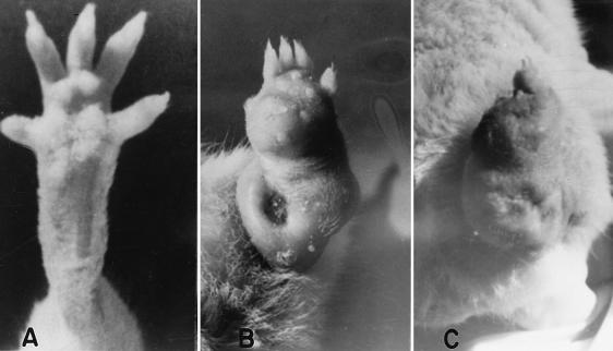
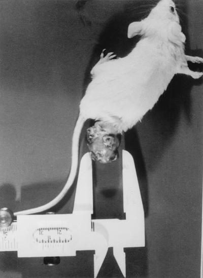
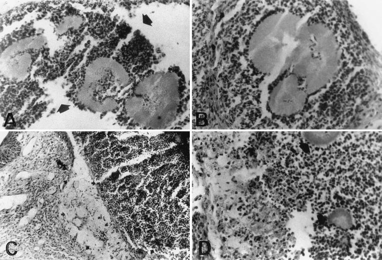
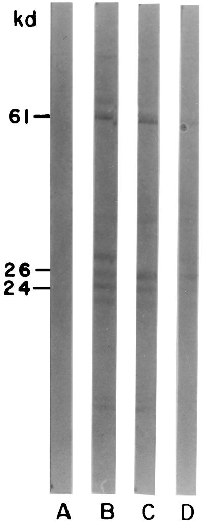
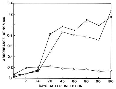
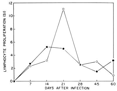
Nine- to twelve-week-old BALB/c mice were injected in footpads with 10(7) CFU of a Nocardia brasiliensis cell suspension. Typical actinomycetoma lesions, characterized by severe local inflammation with abscess and fistula formation, were fully established by day 28 after infection. These changes presented for 90 days, and then tissue repair with scar formation slowly appeared, with complete healing after 150 days of infection. Some animals developed bone destruction in the affected area. Histopathology showed an intense inflammatory response, with polymorphonuclear cells and hyaloid material around the colonies of the bacteria, some of which were discharged from draining abscesses. Sera from experimental animals were analyzed by Western blotting, and immunodominant antigens P61 and P24 were found as major targets for antibody response. Anti-P24 immunoglobulin M (IgM) isotype antibodies were present as early as 7 days, IgG peaking 45 days after infection. Lymphocyte proliferation with spleen and popliteal lymph node cells demonstrated thymidine incorporation at 7 days after infection, the stimulation index decreasing by day 60. Levels of interleukin-1 (IL-1), IL-2, IL-4, IL-6, tumor necrosis factor alpha, and gamma interferon (IFN-gamma) were determined by enzyme-linked immunosorbent assay in the sera of infected animals. The circulating levels of IFN-gamma increased more than 10 times the basal levels; levels of IL-4, IL-6 and IL-10 also increased during the first 4 days of infection.
Figures
Similar articles
-
Humoral immunity through immunoglobulin M protects mice from an experimental actinomycetoma infection by Nocardia brasiliensis.Infect Immun. 2004 Oct;72(10):5597-604. doi: 10.1128/IAI.72.10.5597-5604.2004. Infect Immun. 2004. PMID: 15385456 Free PMC article.
-
IgM but not IgG monoclonal anti-Nocardia brasiliensis antibodies confer protection against experimental actinomycetoma in BALB/c mice.FEMS Immunol Med Microbiol. 2009 Oct;57(1):17-24. doi: 10.1111/j.1574-695X.2009.00575.x. Epub 2009 Jun 10. FEMS Immunol Med Microbiol. 2009. PMID: 19624737
-
Monoclonal antibodies to P24 and P61 immunodominant antigens from Nocardia brasiliensis.Clin Diagn Lab Immunol. 1997 Mar;4(2):133-7. doi: 10.1128/cdli.4.2.133-137.1997. Clin Diagn Lab Immunol. 1997. PMID: 9067645 Free PMC article.
-
Immune response to Nocardia brasiliensis extracellular antigens in patients with mycetoma.Mycopathologia. 2008 Mar;165(3):127-34. doi: 10.1007/s11046-008-9093-4. Epub 2008 Feb 27. Mycopathologia. 2008. PMID: 18302006
-
Nocardia brasiliensis: from microbe to human and experimental infections.Microbes Infect. 2000 Sep;2(11):1373-81. doi: 10.1016/s1286-4579(00)01291-0. Microbes Infect. 2000. PMID: 11018454 Review.
Cited by
-
Nocardia brasiliensis induces an immunosuppressive microenvironment that favors chronic infection in BALB/c mice.Infect Immun. 2012 Jul;80(7):2493-9. doi: 10.1128/IAI.06307-11. Epub 2012 Apr 30. Infect Immun. 2012. PMID: 22547544 Free PMC article.
-
Immunomodulation of IL-1, IL-6 and IL-8 cytokines by Prosopis juliflora alkaloids during bovine sub-clinical mastitis.3 Biotech. 2018 Oct;8(10):409. doi: 10.1007/s13205-018-1438-1. Epub 2018 Sep 14. 3 Biotech. 2018. PMID: 30237956 Free PMC article.
-
A Polymorphism in the Chitotriosidase Gene Associated with Risk of Mycetoma Due to Madurella mycetomatis Mycetoma--A Retrospective Study.PLoS Negl Trop Dis. 2015 Sep 2;9(9):e0004061. doi: 10.1371/journal.pntd.0004061. eCollection 2015. PLoS Negl Trop Dis. 2015. PMID: 26332238 Free PMC article.
-
Nocardia farcinica activates human dendritic cells and induces secretion of interleukin-23 (IL-23) rather than IL-12p70.Infect Immun. 2012 Dec;80(12):4195-202. doi: 10.1128/IAI.00741-12. Epub 2012 Sep 17. Infect Immun. 2012. PMID: 22988018 Free PMC article.
-
Activity of cyclooxygenase-2 and nitric oxide in milk leucocytes following intramammary inoculation of a bio-response modifier during bovine Staphylococcus aureus subclinical mastitis.Vet Res Commun. 2014 Sep;38(3):201-7. doi: 10.1007/s11259-014-9604-3. Epub 2014 May 21. Vet Res Commun. 2014. PMID: 24844929
References
-
- Allen J E, Mizels R M. Th1-Th2: reliable paradigm or dangerous dogma. Immunol Today. 1997;18:387–392. - PubMed
-
- Cremer G, Mezence Baquero M I, Boiron P, Huerre M, Houin R. Experimental mycetoma due to Madurella mycetomatis in non-immunodeficient laboratory mice. Med Microbiol Lett. 1995;4:76–82.
Publication types
MeSH terms
Substances
LinkOut - more resources
Full Text Sources
Molecular Biology Databases
