. 1999 May;67(5):2540-6.
doi: 10.1128/IAI.67.5.2540-2546.1999.

Lipopolysaccharide induction of tissue factor expression in rabbits

Affiliations

Lipopolysaccharide induction of tissue factor expression in rabbits

J Erlich et al. Infect Immun. 1999 May.

Abstract

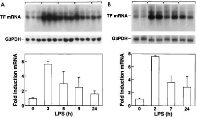
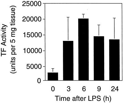
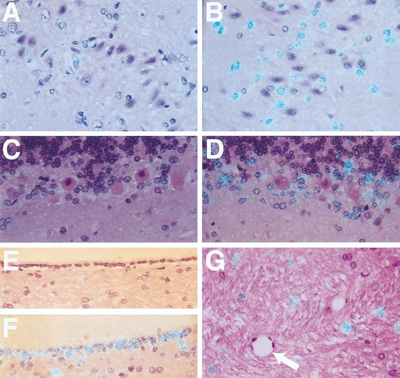
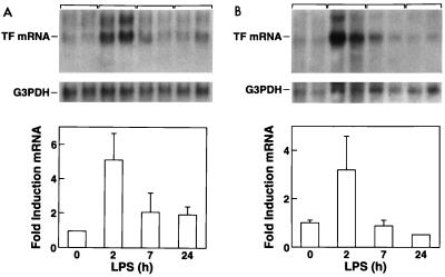
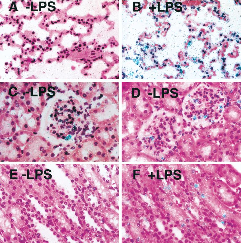
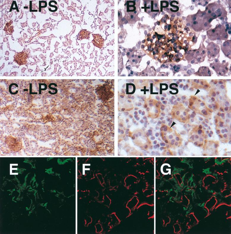
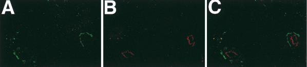
Tissue factor (TF) is the major activator of the coagulation protease cascade and contributes to lethality in sepsis. Despite several studies analyzing TF expression in animal models of endotoxemia, there remains debate about the cell types that are induced to express TF in different tissues. In this study, we performed a detailed analysis of the induction of TF mRNA and protein expression in two rabbit models of endotoxemia to better understand the cell types that may contribute to local fibrin deposition and disseminated intravascular coagulation. Northern blot analysis demonstrated that lipopolysaccharide (LPS) increased TF expression in the brain, lung, and kidney. In situ hybridization showed that TF mRNA expression was increased in cells identified morphologically as epithelial cells in the lung and as astrocytes in the brain. In the kidney, in situ hybridization experiments and immunohistochemical analysis showed that TF mRNA and protein expression was increased in renal glomeruli and induced in tubular epithelium. Dual staining for TF and vWF failed to demonstrate TF expression in endothelial cells in LPS-treated animals. These results demonstrate that TF expression is induced in many different cell types in LPS-treated rabbits, which may contribute to local fibrin deposition and tissue injury during endotoxemia.


Figures

FIG. 1
FIG. 1
LPS induction of TF mRNA in the brain. (A) TF mRNA expression in a three-injection model. Tissues were collected 3, 6, 9, and 24 h after the last dose of LPS. Quantitation of brain TF mRNA was performed by densitometric analysis of the Northern blots. TF mRNA levels were normalized by using G3PDH mRNA levels and expressed as fold induction ± SD. (B) TF mRNA expression in a single-injection model.
FIG. 2
FIG. 2
LPS induction of TF activity in the brain. Brain extracts were prepared from control and LPS-treated rabbits (three-injection model), and TF procoagulant activity was measured by a single-stage clotting assay. Data are presented as means ± SD.
FIG. 3
FIG. 3
Localization of LPS-inducible TF mRNA in the brain. In situ hybridization was performed on brain sections from control rabbits (A, C, and E) and from rabbits 3 h after the last LPS injection in a three-injection model (B, D, F, and G). The cerebral cortex (A and B), cerebellum (C and D), ependymal cell lining of the ventricles (E and F), and brain stem (G) are shown (the arrow indicates a blood vessel). All sections were hybridized with a radiolabeled antisense TF probe (magnification, ×368). Exposure time was 8 weeks.
FIG. 4
FIG. 4
LPS induction of TF mRNA expression in the lung (A) and kidney (B). Northern blot analysis of TF mRNA and G3PDH mRNA from rabbits treated for different times with a single 10-μg dose of LPS. TF mRNA levels were normalized with G3PDH and expressed as fold induction ± SD.
FIG. 5
FIG. 5
Localization of LPS-inducible TF mRNA expression in lung and kidney. In situ hybridization for TF mRNA was performed on pulmonary and renal tissue collected from control rabbits (A, C, and E) and rabbits 2 h after treatment with a single dose of LPS (B, D, and F). The lung (A and B), glomeruli (C and D), and renal tubules (E and F) are shown. All sections were hybridized with a radiolabeled TF antisense probe (magnification, ×332). The exposure time was 8 weeks.
FIG. 6
FIG. 6
Immunolocalization of TF in the kidney after administration of a single dose of LPS. Sections from control rabbits (A and B) and rabbits 7 h after a single dose of LPS (C and D) are shown. The sections were photographed at ×82 (A and C) or ×328 (B and D) magnification. The arrowheads (D) indicate TF-positive tubular cells. No staining was observed with a control antibody (data not shown). (E, F, and G) Immunofluorescence staining detected by confocal microscopy for TF (11F) and vWF (goat anti-rabbit vWF antibody) on histological sections from a rabbit 24 h after a single 10-μg dose of LPS. Green staining (TF) is observed in the mesangium but is not found on the endothelium of the glomerular capillary tuft. Red staining (vWF) marks the endothelium. (E) TF staining alone, (F) vWF staining alone; (G) combined confocal image of TF and vWF staining (magnification, ×328).
FIG. 7
FIG. 7
TF expression in the spleen. Seven hours after a single dose of LPS, green-staining TF is observed on adventitial cells of the microvasculature but is not found on endothelial cells. Red staining (vWF) marks the endothelium. (A) TF staining alone; (B) vWF staining alone; (C) combined confocal image of TF and vWF staining (magnification, ×400).

References

    1. Andrews B S, Rehemtulla A, Fowler B J, Edgington T S, Mackman N. Conservation of tissue factor primary sequence among three mammalian species. Gene. 1991;98:265–269. - PubMed
    1. Bach R R. Initiation of coagulation by tissue factor. Crit Rev Biochem. 1988;23:339–368. - PubMed
    1. Broze G J, Leyham J E, Schwartz B D, Miletich J P. Purification of human brain tissue factor. J Biol Chem. 1985;260:10917–10920. - PubMed
    1. Brukman J, Wiggins R C. Procoagulant activity in kidneys of normal and bacterial lipopolysaccharide-treated rabbits. Kidney Int. 1987;32:31–38. - PubMed
    1. Chomczynski P, Sacchi N. Single-step method of RNA isolation by acid guanidinium thiocyanate-phenol-chloroform extraction. Anal Biochem. 1987;162:156–159. - PubMed

Publication types

LinkOut - more resources