. 1999 May;67(5):2567-74.
doi: 10.1128/IAI.67.5.2567-2574.1999.

Yersinia pseudotuberculosis-induced calcium signaling in neutrophils is blocked by the virulence effector YopH

Affiliations

Yersinia pseudotuberculosis-induced calcium signaling in neutrophils is blocked by the virulence effector YopH

K Andersson et al. Infect Immun. 1999 May.

Abstract

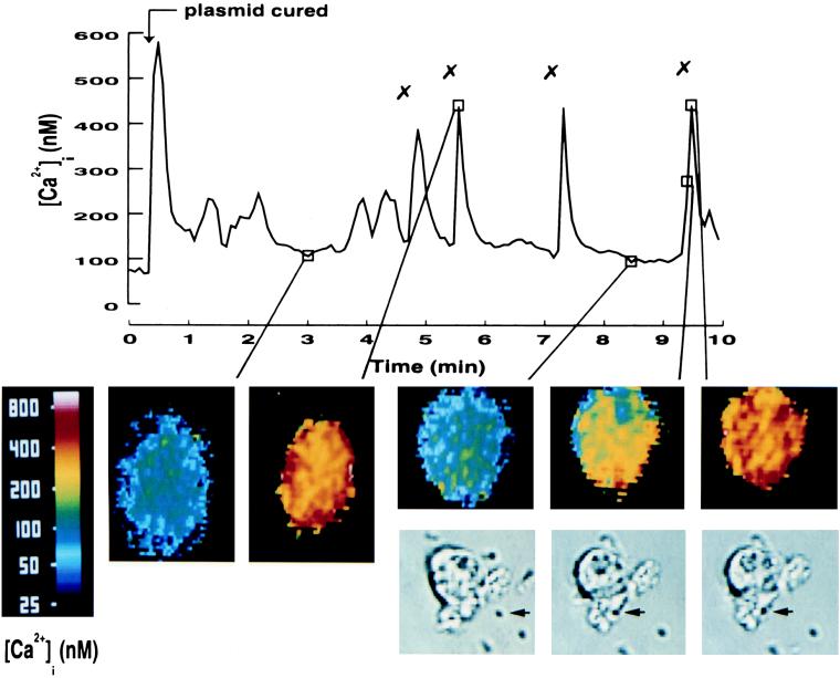
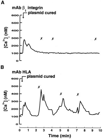
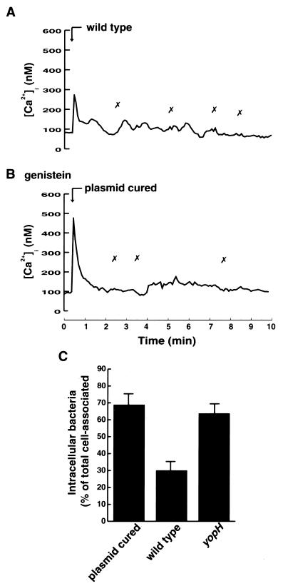
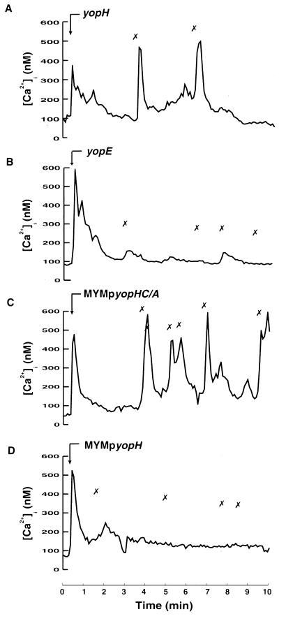
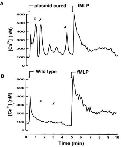
Pathogenic species of the genus Yersinia evade the bactericidal functions of phagocytes. This evasion is mediated through their virulence effectors, Yops, which act within target cells. In this study we investigated the effect of Yersinia pseudotuberculosis on Ca2+ signaling in polymorphonuclear neutrophils. The intracellular free calcium concentration in single adherent human neutrophils was monitored during bacterial infection and, in parallel, the encounter between the bacteria and cells was observed. When a plasmid-cured strain was used for infection, adherence of a single bacterium to the cellular surface induced a beta1 integrin-dependent transient increase in the intracellular concentration of free calcium. This was, however, not seen with Yop-expressing wild-type bacteria, which adhered to the cell surface without generating any Ca2+ signal. Importantly, the overall Ca2+ homeostasis was not affected by the wild-type strain; the Ca2+ signal mediated by the G-protein-coupled formyl-methionyl-leucyl-phenylalanine receptor was still functioning. Hence, the blocking effect was restricted to certain receptors and their signaling pathways. The use of different Yop mutant strains revealed that the protein tyrosine phosphatase YopH was responsible for the inhibition. This virulence determinant has previously been implicated in very rapid Yersinia-mediated effects on target cells as the key effector in the blockage of phagocytic uptake. The present finding, that Y. pseudotuberculosis, via YopH, specifically inhibits a self-induced immediate-early Ca2+ signal in neutrophils, offers more-detailed information concerning the effectiveness of this virulence effector and implies an effect on Ca2+-dependent, downstream signals.


Figures

FIG. 1
FIG. 1
Bacterium-induced Ca2+ signaling in human neutrophils. Time course of [Ca2+]i in a single neutrophil upon infection with Y. pseudotuberculosis plasmid-cured bacteria. The plasmid-cured strain was added to adherent neutrophils at a calculated bacterium/cell ratio of 50:1 (arrow). The “✗” symbol indicates contact between a bacterium and the neutrophil. Attachment of bacteria was visually observed on a video screen, and this was correlated to the [Ca2+]i transients. A representative time course is presented (of 12 separate experiments), together with some selected images in pseudocolor (time points are indicated in the graph). The phase images show the attachment of a single bacterium (indicated by an arrow) to the same neutrophil shown in the pseudocolor image situated above. The mean number of [Ca2+]i transients is shown in Table 2.
FIG. 2
FIG. 2
Involvement of β1 integrins in Yersinia-induced [Ca2+]i elevations in neutrophils. Time courses of [Ca2+]i in single neutrophils after receptor blockage and during infection with Y. pseudotuberculosis are shown. Adherent neutrophils were pretreated with blocking antibodies (10 μg/ml) directed against β1 integrins (A) or HLA (B) for 10 min. The Y. pseudotuberculosis plasmid-cured strain was then added at a calculated bacterium/cell ratio of 50:1 (arrow). The “✗” symbol indicates contact between a bacterium and the neutrophil. Attachment of bacteria was visually observed on a video screen, and this was correlated with the [Ca2+]i transients. Representative time courses are presented (of 4 to 10 separate experiments). The mean numbers of [Ca2+]i transients are shown in Table 2.
FIG. 3
FIG. 3
Inhibition of bacterium-induced elevations in [Ca2+]i. (A and B) Time courses of [Ca2+]i in single neutrophils upon infection with Y. pseudotuberculosis wild-type strain (A) or plasmid-cured strain (B) at a calculated bacterium/cell ratio of 50:1 (arrow). In panel B, the neutrophils were preincubated at 37°C for 15 min with the tyrosine kinase inhibitor genistein before bacterial infection. The “✗” symbols indicate contact between bacteria and the neutrophil. Attachment of bacteria was visually observed on a video screen, and this was correlated with the [Ca2+]i transients. Representative time courses are presented (of 9 to 13 separate experiments). The mean number of [Ca2+]i transients is shown in Table 2. (C) Phagocytosis of Y. pseudotuberculosis strains. The plasmid-cured strain, the wild type, and the yopH mutant were exposed to neutrophils at a calculated bacterium/cell ratio of 50:1 for 30 min. Determinations of cellular association and discrimination between intra- and extracellularly associated bacteria were done as described in Materials and Methods. The data given represent means ± the standard errors of the means of five separate experiments and are expressed as percentages of neutrophil-associated bacteria that were located intracellularly.
FIG. 4
FIG. 4
Involvement of YopH in inhibition of intracellular Ca2+ signaling. Time courses of [Ca2+]i in single neutrophils during attachment of different Y. pseudotuberculosis strains are shown. Adherent neutrophils were exposed to the Yersinia yopH mutant (A), yopE mutant (B), MYM transcomplemented with yopHC/A (C), or MYM transcomplemented with wild-type yopH (D) at a calculated bacterium/cell ratio of 50:1 (arrow). The “✗” symbols indicate contact between the bacteria and the neutrophil. Attachment of bacteria was visually observed on a video screen, and this was correlated with the [Ca2+]i transients. Representative time courses are presented (of 9 to 12 separate experiments). The mean numbers of [Ca2+]i transients are shown in Table 2.
FIG. 5
FIG. 5
Receptor specificity of the wild-type-mediated inhibition. Time courses of [Ca2+]i in single neutrophils during infection with Y. pseudotuberculosis and subsequent stimulation with fMLP are shown. Adherent neutrophils were exposed to Yersinia plasmid-cured strain (A) or to the wild-type strain (B) at a calculated bacterium/cell ratio of 50:1 for 5 min. The “✗” symbols indicate contact between bacteria and the neutrophil. Neutrophils were then stimulated with 10−8 M fMLP (arrow). Representative time courses are presented (of three separate experiments).

References

    1. Andersson K, Carballeira N, Magnusson K-E, Persson C, Stendahl O, Wolf-Watz H, Fällman M. YopH of Yersinia pseudotuberculosis interrupts early phosphotyrosine signalling associated with phagocytosis. Mol Microbiol. 1996;20:1057–1069. - PubMed
    1. Aplin A, Howe A, Alahari S, Juliano R. Signal transduction and signal modulation by cell adhesion receptors: the role of integrins, cadherins, immunoglobulin-cell adhesion molecules, and selectins. Pharmacol Rev. 1998;50:197–263. - PubMed
    1. Bei L, Hu T, Qian Z, Shen X. Extracellular Ca2+ regulates the respiratory burst of human neutrophils. Biochim Biophys Acta. 1998;1404:475–483. - PubMed
    1. Black D S, Bliska J B. Identification of p130Cas as a substrate of Yersinia YopH (Yop51), a bacterial protein tyrosine phosphatase that translocates into mammalian cells and targets focal adhesions. EMBO J. 1997;16:2730–2744. - PMC - PubMed
    1. Bliska J B, Black D S. Inhibition of the Fc receptor-mediated oxidative burst in macrophages by the Yersinia pseudotuberculosis tyrosine phosphatase. Infect Immun. 1995;63:681–685. - PMC - PubMed

Publication types

MeSH terms

LinkOut - more resources