. 1999 May 3;145(3):437-45.
doi: 10.1083/jcb.145.3.437.

Light-dependent translocation of a phytochrome B-GFP fusion protein to the nucleus in transgenic Arabidopsis

Affiliations

Light-dependent translocation of a phytochrome B-GFP fusion protein to the nucleus in transgenic Arabidopsis

R Yamaguchi et al. J Cell Biol. .

Abstract

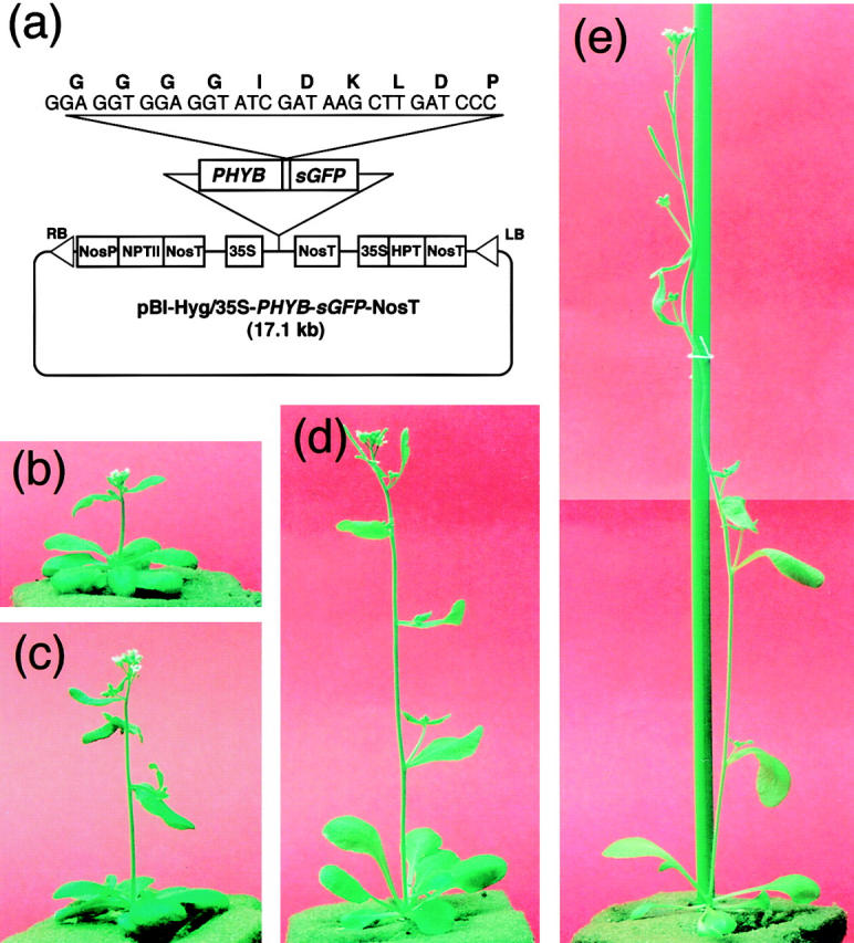
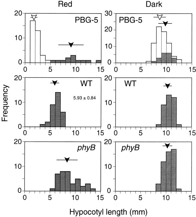
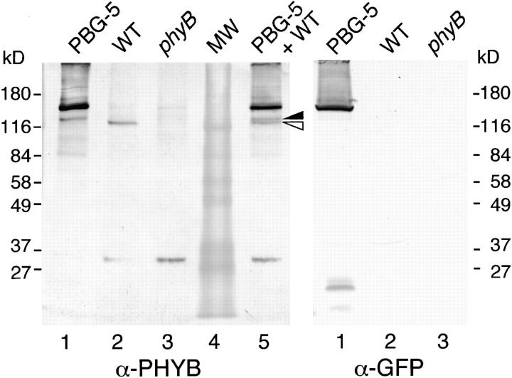
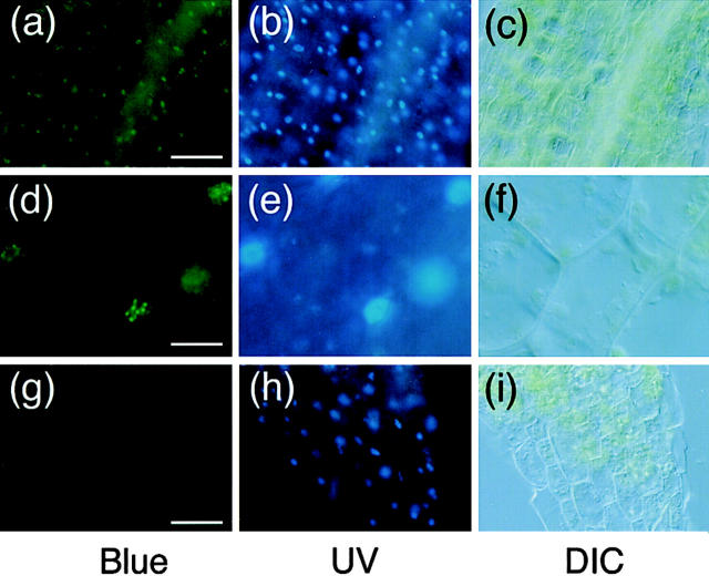
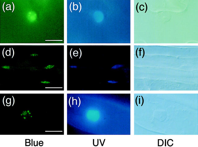
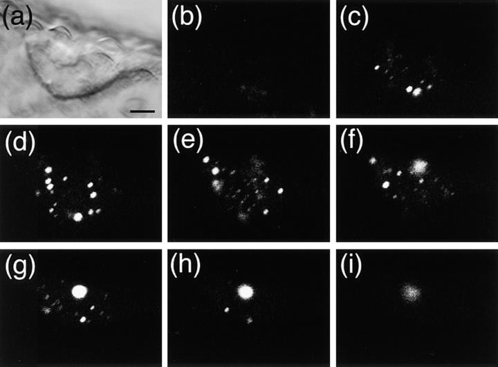
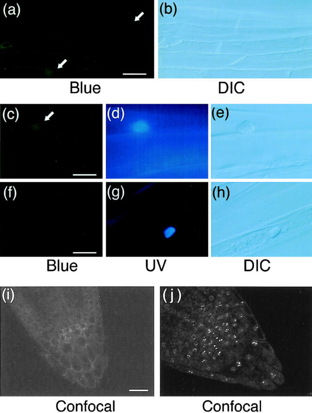
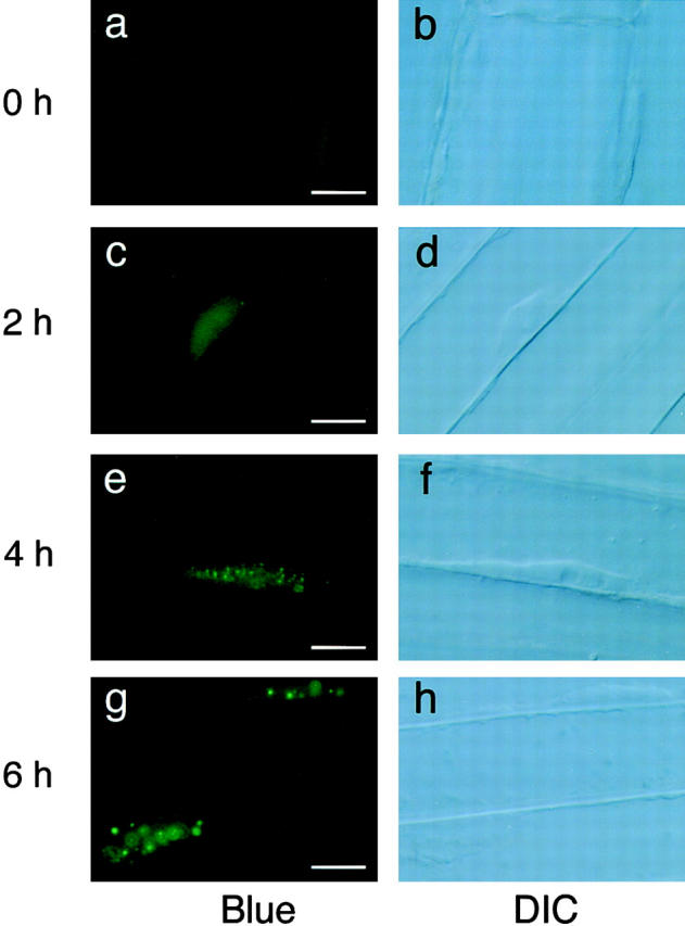
Phytochrome is a ubiquitous photoreceptor of plants and is encoded by a small multigene family. We have shown recently that a functional nuclear localization signal may reside within the COOH-terminal region of a major member of the family, phytochrome B (phyB) (Sakamoto, K., and A. Nagatani. 1996. Plant J. 10:859-868). In the present study, a fusion protein consisting of full-length phyB and the green fluorescent protein (GFP) was overexpressed in the phyB mutant of Arabidopsis to examine subcellular localization of phyB in intact tissues. The resulting transgenic lines exhibited pleiotropic phenotypes reported previously for phyB overexpressing plants, suggesting that the fusion protein is biologically active. Immunoblot analysis with anti-phyB and anti-GFP monoclonal antibodies confirmed that the fusion protein accumulated to high levels in these lines. Fluorescence microscopy of the seedlings revealed that the phyB-GFP fusion protein was localized to the nucleus in light grown tissues. Interestingly, the fusion protein formed speckles in the nucleus. Analysis of confocal optical sections confirmed that the speckles were distributed within the nucleus. In contrast, phyB-GFP fluorescence was observed throughout the cell in dark-grown seedlings. Therefore, phyB translocates to specific sites within the nucleus upon photoreceptor activation.


Figures

Figure 1
Figure 1
Two independent lines of transgenic Arabidopsis, PBG-5 and PBG-7, which overexpress the phyB-GFP fusion protein. Plants were grown for 4 wk under continuous white light. (a) pBI-Hyg/35S-PHYB-sGFP-NosT used for transformation of Arabidopsis plants. sGFP, synthetic GFP; RB, right border of T-DNA; LB, left border of T-DNA; NosP, nopaline synthase promoter; NosT, nopaline synthase terminator; NPTII, neomycin phosphotransferase II; 35S, cauliflower mosaic virus 35S promoter; HPT, hygromycin phosphotransferase. (b) Picture of the PBG-7 plant. (c) Picture of the PBG-5 plant. (d) Picture of the wild-type plant. (e) Picture of the phyB-5 mutant plant.
Figure 2
Figure 2
Frequency distribution of hypocotyl lengths in PBG-5, phyB mutant, and the wild-type seedlings under continuous red light (left) or in darkness (right). Heterozygous progeny of PBG-5 plant was examined. Individuals that exhibited GFP fluorescence are unshaded. Open and closed arrowheads indicate average hypocotyl lengths of fluorescent and nonfluorescent populations, respectively. Bars indicate the standard deviation.
Figure 3
Figure 3
Immunoblot analysis of phyB-GFP fusion protein in the PBG-5 rosette leaves. Extracts from rosette leaves were probed with anti-phyB (left) or anti-GFP mAb (right). Lane 1, PBG-5; lane 2, the wild-type; lane 3, the phyB mutant; lane 4, molecular weight markers; lane 5, 1:1 mixture of the extracts from PBG-5 and the wild-type plants. Closed triangle, a proteolytic phyB-GFP fragment; open triangle, authentic phyB. Each lane contains either 25 (left) or 112 (right) μg total protein.
Figure 4
Figure 4
Fluorescence microscopic observation of hypocotyl peel from light-grown PBG-5 and the wild-type seedlings. Samples were stained with Hoechst No. 33342 and viewed under epifluorescence optics with blue (left) or UV (middle) excitation. DIC images in the same view are shown (right). (a–c) PBG-5 hypocotyl cells, ×20 objective. Bar, 50 μm. (d–f) PBG-5 hypocotyl cells, ×100 objective. Bar, 10 μm. (g–i) Wild-type hypocotyl cells, ×20 objective. Bar, 50 μm.
Figure 5
Figure 5
Fluorescence microscopic images of different parts of light-grown PBG-5 seedlings. Samples were stained with Hoechst No. 33342 and viewed under epifluorescence optics with blue (left) and UV (middle) excitation. DIC images of the same sample are shown (right). (a–c) PBG-5 leaf epidermis, ×100 objective. Bar, 10 μm. (d–f) PBG-5 root cells, ×40 objective. Bar, 25 μm. (g–i) PBG-root hair, ×100 objective. Bar, 10 μm.
Figure 6
Figure 6
Confocal optical sectioning of the trichome nucleus in PBG-5. Trichomes were removed from light-grown PBG-5 cotyledons and observed on an inverted laser scan microscope (LSM410 invert; Carl Zeiss Jena) with a combination of 488 nm laser excitation and 515 nm longpass emission filter. (a) DIC image, ×63 objective. Bar, 5 μm. (b–i) Serial sections at 2-μm intervals, ×63 objective.
Figure 7
Figure 7
Fluorescence microscopic images of hypocotyl and root tip cells in dark-grown PBG-5 and wild-type seedlings. Hypocotyl specimens were stained with Hoechst No. 33342 and viewed under epifluorescence optics with blue (a, c, and f) and UV (d and g) excitation or under DIC optics (b, e, and h). Root tip specimens were observed on an inverted laser scan microscope (LSM410 invert; Carl Zeiss Jena) with a combination of 488 nm laser excitation and 515 nm longpass emission filter (i and j). Arrows indicate fluorescence detected in the nuclear regions. (a and b) Dark-grown PBG-5 hypocotyl cells, ×40 objective. Bar, 25 μm. (c–e) Dark-grown PBG-5 hypocotyl cells, ×100 objective. Bar, 10 μm. (f–h) Dark-grown wild-type hypocotyl cells, ×100 objective. Bar, 10 μm. (i) Dark-grown PBG-5 root tip cells, ×40 objective. Bar, 20 μm. (j) Light-grown PBG-5 root tip cells, ×40 objective.
Figure 8
Figure 8
Fluorescence microscopic images of hypocotyl cells in PBG-5 dark-grown seedlings placed under continuous red light for different duration. Hypocotyl cells were viewed under epifluorescence optics with blue excitation (left) and DIC optics (right) with a ×100 objective. Bar, 10 μm. (a and b) Dark-grown seedling. (c and d) 2 h in red light. (e and f) 4 h in red light. (g and h) 6 h in red light.

References

    1. Anderson SL, Somers DE, Millar AJ, Hanson K, Chory J, Kay SA. Attenuation of phytochrome A and B signaling pathways by the Arabidopsiscircadian clock. Plant Cell. 1997;9:1727–1743. - PMC - PubMed
    1. Ang L-H, Chattopadhyay S, Wei N, Oyama T, Okada K, Batschauer A, Deng X-W. Molecular interaction between COP1 and HY5 defines a regulatory switch for light control of Arabidopsisdevelopment. Mol Cell. 1998;1:213–222. - PubMed
    1. Aukerman MJ, Hirschfeld M, Wester L, Weaver M, Clack T, Amasino RM, Sharrock RA. A deletion in the PHYD gene of the ArabidopsisWassilewskija ecotype defines a role for phytochrome D in red/far-red light sensing. Plant Cell. 1997;9:1317–1326. - PMC - PubMed
    1. Bechtold N, Ellis J, Pelletier G. In planta Agrobacterium mediated gene transfer by infiltration of adult Arabidopsis thalianaplants. CR Acad Sci Paris Life Sci. 1993;316:1194–1199.
    1. Chattopadhyay S, Ang L-H, Puente P, Deng X-W, Wei N. ArabidopsisbZIP protein HY5 directly interacts with light-responsive promoters in mediating light control of gene expression. Plant Cell. 1998;10:673–684. - PMC - PubMed

Publication types

MeSH terms