. 1999 May 3;145(3):457-68.
doi: 10.1083/jcb.145.3.457.

Trafficking of an acylated cytosolic protein: newly synthesized p56(lck) travels to the plasma membrane via the exocytic pathway

Affiliations

Trafficking of an acylated cytosolic protein: newly synthesized p56(lck) travels to the plasma membrane via the exocytic pathway

M J Bijlmakers et al. J Cell Biol. .

Abstract

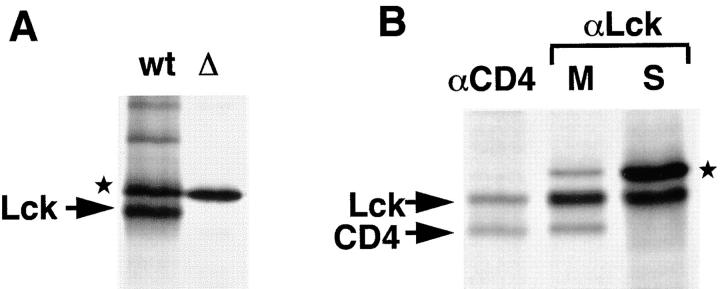
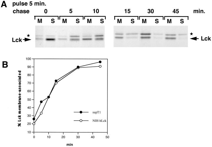
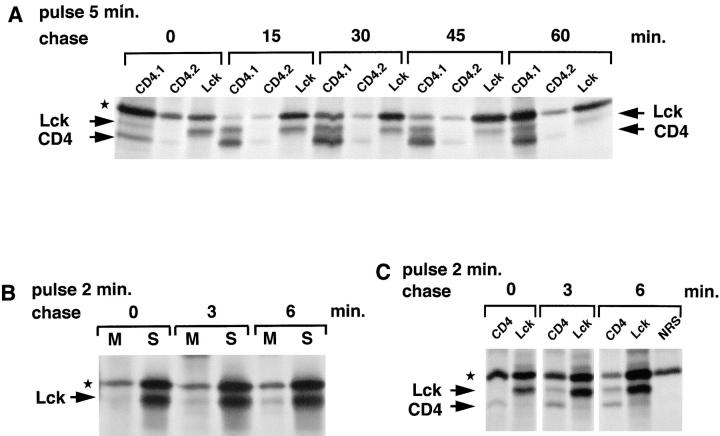
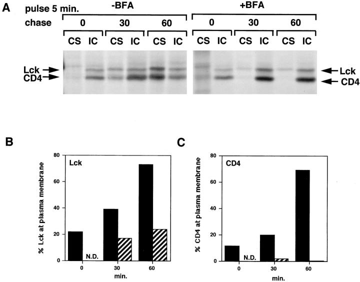
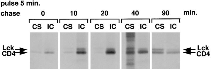
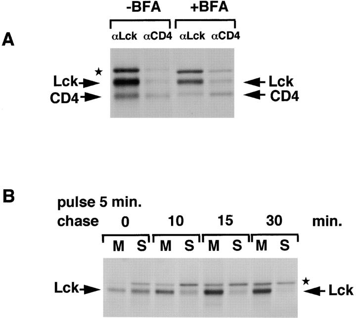
The Src-related tyrosine kinase p56(lck) (Lck) is primarily expressed in T lymphocytes where it localizes to the cytosolic side of the plasma membrane and associates with the T cell coreceptors CD4 and CD8. As a model for acylated proteins, we studied how this localization of Lck is achieved. We followed newly synthesized Lck by pulse-chase analysis and found that membrane association of Lck starts soon after synthesis, but is not complete until at least 30-45 min later. Membrane-binding kinetics are similar in CD4/CD8-positive and CD4/CD8-negative cells. In CD4-positive T cells, the interaction with CD4 rapidly follows membrane association of Lck. Studying the route via which Lck travels from its site of synthesis to the plasma membrane, we found that: CD4 associates with Lck within 10 min of synthesis, long before CD4 has reached the plasma membrane; Lck associates with intracellular CD4 early after synthesis and with cell surface CD4 at later times; and transport of CD4-bound Lck to the plasma membrane is inhibited by Brefeldin A. These data indicate that the initial association of newly synthesized Lck with CD4, and therefore with membranes, occurs on intracellular membranes of the exocytic pathway. From this location Lck is transported to the plasma membrane.


Figures

Figure 1
Figure 1
Characterization of Lck antiserum. (A) Jurkat (wt) and mutant JCam.1 (Δ) cells (6 × 10⁶ each) were labeled for 30 min with [35S]methionine/cysteine (0.5 mCi) and subjected to immunoprecipitation with LckN serum. JCam.1 does not express wt Lck (Straus and Weiss, 1992). In addition to Lck, the LckN antiserum recognizes a protein with an apparent molecular mass of 60 kD (★). (B) SupT1 cells were labeled with [35S]methionine/ cysteine for 5 min and chased for 15 min. Cells were broken in hypotonic buffer, nuclei removed by centrifugation, and membranes recovered by centrifugation at 100,000 g for 45 min. Membrane and soluble fractions were split into two: half was subjected to immunoprecipitation with anti-CD4 antibodies (αCD4), the other half with anti-Lck antibodies (αLck). The CD4 immunoprecipitation from membranes, and the Lck immunoprecipitation both from the membrane (M) and soluble (S) fractions are shown. CD4, Lck, and the background band (★) are indicated.
Figure 2
Figure 2
Membrane-binding kinetics of Lck. (A) SupT1 cells were broken in hypotonic buffer and Dounce homogenized. After removal of nuclei (5 min at 1,500 rpm), membrane and soluble fractions were separated by centrifugation at 100,000 g. Equivalent amounts of total (T), nuclear (N), membrane (M), and soluble (S) fractions were analyzed by immunoblotting with LckC. (B) SupT1 cells (2 × 10⁸) were labeled with [35S]methionine/cysteine (2 mCi) for 5 min and chased for the indicated times. Lck was immunoprecipitated from the membrane (M) and soluble (S) fractions with LckN and analyzed by SDS-PAGE and autoradiography. Lck and CD4 are indicated. ★ indicates the background band.
Figure 3
Figure 3
Lck membrane-binding kinetics in the presence and absence of CD4. (A) CD4-negative Jurkat cells were labeled for 5 min and chased as indicated. Lck was immunoprecipitated from membrane (M) and soluble (S) fractions as described for Fig. 2 B. ★ indicates the background band. (B) The same experiments as in A, now for CD4-positive Jurkat cells. (C) Cell lysates of CD4-negative (−) and CD4-positive (+) Jurkat cells were analyzed for CD4 expression by immunoblotting. (D) Quantitation of Lck membrane-binding experiments for two CD4-negative and one CD4-positive Jurkat cell line. Autoradiograms were digitized (as described in Materials and Methods) and analyzed using the Molecular Analyst program (Bio-Rad). Membrane-associated Lck is expressed as a percentage of the total amount of Lck and plotted against time.
Figure 4
Figure 4
Lck membrane-binding kinetics in NIH-3T3 cells. (A) NIH-3T3 cells stably transfected with Lck were pulse-labeled and chased as indicated and Lck was immunoprecipitated from the membrane (M) and soluble (S) fractions. ★ indicates the background band. (B) Comparison of Lck membrane-binding kinetics in SupT1 and NIH-3T3 cells. The experiments shown here and in Fig. 2 B were quantitated. The percentage of membrane-associated Lck is plotted against time.
Figure 5
Figure 5
Kinetics of the association of Lck with CD4. (A) SupT1 cells (1.25 × 10⁸) were labeled for 5 min with [35S]methionine/cysteine (1.5 mCi) and chased as indicated. The cells were lysed in NP-40 buffer and subjected to sequential immunoprecipitations, first with two rounds of anti-CD4 antibodies (CD4.1 and CD4.2) and next with anti-Lck antibodies (Lck). (B) SupT1 cells (108) were labeled with [35S]methionine/cysteine (1 mCi) for 2 min and chased for 0, 3, or 6 min. Lck was immunoprecipitated from membrane (M) and soluble (S) fractions with LckN and analyzed by SDS-PAGE and autoradiography. (C) SupT1 cells (7.5 × 10⁷) were labeled with [35S]methionine/cysteine (1 mCi) for 2 min and chased for 0, 3, or 6 min. Cell lysates were subjected to sequential immunoprecipitations, first with anti-CD4 antibodies (CD4) and next with anti-Lck antibodies (Lck). The last of three rounds of preclears with normal rabbit serum (NRS) and protein A–Sepharose is shown for the 6-min chase (NRS). Lck, CD4, and the background band (★) are indicated.
Figure 8
Figure 8
Transport of CD4-associated Lck is inhibited in the presence of BFA. (A) SupT1 cells were labeled in the absence or presence of BFA. BFA was added 2 min before pulse-labeling at a concentration of 10 μg/ml and was present during the chase at 2 μg/ml. Cell surface (CS) and intracellular (IC) CD4 were immunoprecipitated separately as described for Fig. 7. (B) Quantitation of the autoradiogram in A for Lck. Per time point, the amount of Lck associated with cell surface (CS) CD4 was expressed as a percentage of the total (CS + IC) Lck coimmunoprecipitated with CD4. Black bars, without BFA; striped bars, with BFA. (C) Quantitation of the autoradiogram in A for CD4. Per time point the relative amount of CD4 detected in the cell surface (CS) immunoprecipitation was expressed as a percentage of the total (CS + IC) amount of CD4. Black bars, without BFA; striped bars, with BFA.
Figure 7
Figure 7
Lck associates predominantly with intracellular CD4 early after synthesis. SupT1 cells (1.5 × 10⁸) were labeled with [35S]methionine/cysteine (1.5 mCi) for 5 min and chased for the indicated times. The intact cells were incubated with antibodies against CD4 for 1 h at 4°C. After extensive washing, the cells were lysed in the presence of soluble CD4 to block free antibody-binding sites, and cell surface CD4 (CS) was recovered by incubation with protein A–Sepharose. Subsequently, anti-CD4 antibodies were added to the lysates to recover intracellular CD4 (IC). CD4 and coimmunoprecipitated Lck are indicated.
Figure 6
Figure 6
Kinetics of the association of CD4 with Lck. (A) SupT1 cells (5 × 10⁷) were labeled for 5 min with [35S]methionine/cysteine (0.5 mCi) and chased for the indicated times. The cells were lysed in NP-40 buffer and subjected to immunoprecipitation with anti-LckN serum. Association of CD4 with Lck was detected by coimmunoprecipitation with Lck. Lck, CD4, and the background band (★) are indicated. (B) SupT1 cells were labeled as described in A, lysed, and CD4 was immunoprecipitated from the cell lysates. Immunoprecipitates were incubated with Endo H (1 mU) for 1 h at 37°C before separation by SDS-PAGE. Endo H–sensitive (S) forms of CD4 from which two carbohydrate chains were removed and resistant (R) forms from which only one carbohydrate chain was removed are indicated. Nondigested CD4, containing two carbohydrate chains, migrates slower than Endo H–resistant CD4 (not shown). Coimmunoprecipitated Lck is indicated and is unaffected by Endo H treatment.
Figure 9
Figure 9
Binding of Lck to newly synthesized CD4 and membranes in the presence of BFA. (A) SupT1 cells were pulse-labeled for 5 min and chased for 40 min in the absence or presence of BFA (10 μg/ml during pulse, 2 μg/ml during chase). Pretreatment with BFA was 2 min before the pulse. NP-40 cell lysates were subjected to immunoprecipitation, first with anti-Lck (αLck lanes), next with anti-CD4 antibodies (αCD4). Lck, CD4, and a background band (★) are indicated. (B) SupT1 cells were pretreated and labeled for 5 min in the presence of BFA (10 μg/ml) and chased for indicated times in the presence of 2 μg/ml BFA. Lck was immunoprecipitated at indicated chase times from membrane (M) and soluble (S) fractions.

References

    1. Alland L, Peseckis SM, Atherton RE, Berthiaume L, Resh MD. Dual myristylation and palmitylation of Src family member p59fyn affects subcellular localization. J Biol Chem. 1994;24:16701–16705. - PubMed
    1. Berthiaume L, Resh MD. Biochemical characterization of a palmitoyl acyltransferase activity that palmitoylates myristoylated proteins. J Biol Chem. 1995;270:22399–22405. - PubMed
    1. Bhatnagar RS, Gordon JI. Understanding covalent modifications of proteins by lipids: where cell biology and biophysics mingle. Trends Cell Biol. 1997;7:14–21. - PubMed
    1. Bijlmakers MJE, Isobe-Nakamura M, Ruddock LJ, Marsh M. Intrinsic signals in the unique domain target p56(lck) to the plasma membrane independently of CD4. J Cell Biol. 1997;137:1029–1040. - PMC - PubMed
    1. Bonatti S, Migliaccio G, Simons K. Palmitylation of viral membrane glycoproteins takes place after exit from the endoplasmic reticulum. J Biol Chem. 1989;264:12590–12595. - PubMed

Publication types

MeSH terms