. 1999 May 3;145(3):503-14.
doi: 10.1083/jcb.145.3.503.

Abnormal reaction to central nervous system injury in mice lacking glial fibrillary acidic protein and vimentin

Affiliations

Abnormal reaction to central nervous system injury in mice lacking glial fibrillary acidic protein and vimentin

M Pekny et al. J Cell Biol. .

Abstract

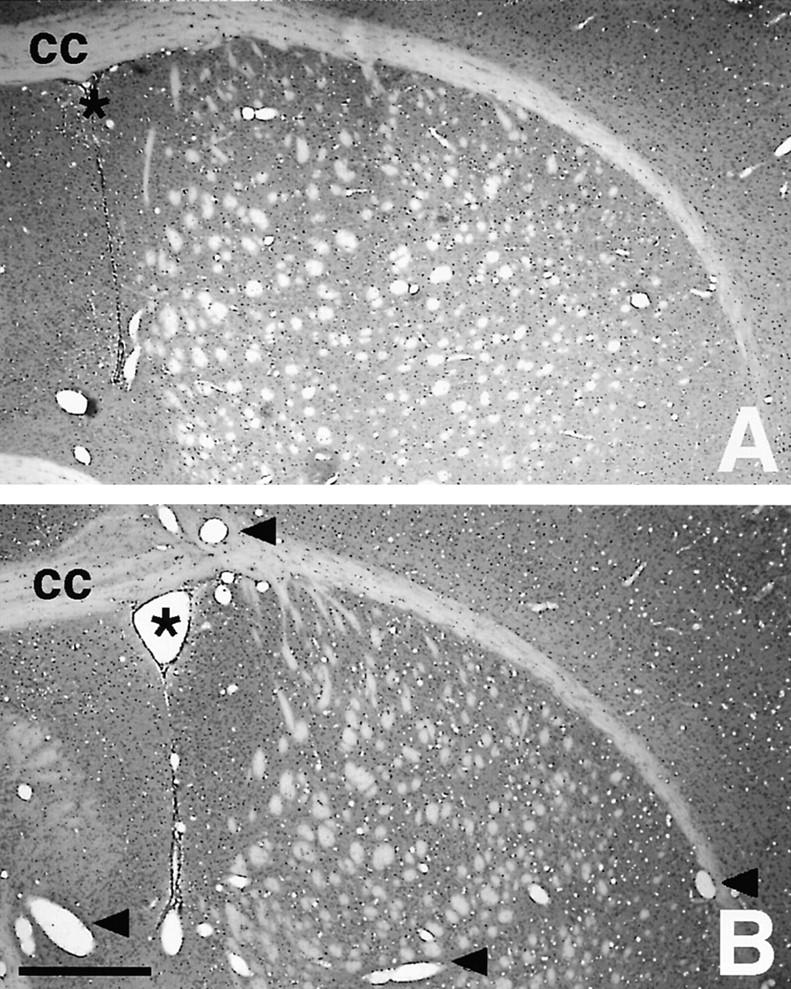
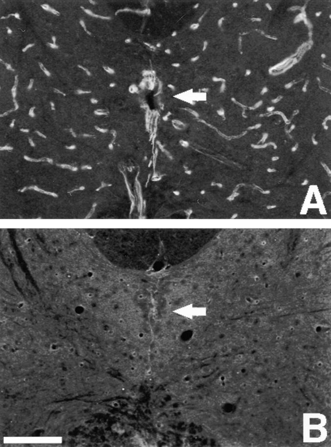
In response to injury of the central nervous system, astrocytes become reactive and express high levels of the intermediate filament (IF) proteins glial fibrillary acidic protein (GFAP), vimentin, and nestin. We have shown that astrocytes in mice deficient for both GFAP and vimentin (GFAP-/-vim-/-) cannot form IFs even when nestin is expressed and are thus devoid of IFs in their reactive state. Here, we have studied the reaction to injury in the central nervous system in GFAP-/-, vimentin-/-, or GFAP-/-vim-/- mice. Glial scar formation appeared normal after spinal cord or brain lesions in GFAP-/- or vimentin-/- mice, but was impaired in GFAP-/-vim-/- mice that developed less dense scars frequently accompanied by bleeding. These results show that GFAP and vimentin are required for proper glial scar formation in the injured central nervous system and that some degree of functional overlap exists between these IF proteins.


Figures

Figure 1
Figure 1
Blood vessel dilation. Hematoxylin and eosin stained coronal brain sections from wild-type (A) and GFAP−/−vim−/− (B) mice show an increased number of large diameter blood vessels in GFAP−/−vim−/− mice (some are indicated by arrowheads in B). The lumen of the lateral ventricle is indicated with an asterisk and the corpus callosum is labeled cc. Bar, 500 μm.
Figure 2
Figure 2
Quantification of blood vessel dilation and the indentation below the dorsal spinal vein. (A) Average number of blood vessels with a diameter greater than 15 μm in spinal cord sections of mice of different genotypes. (B) Depth of the indentation in the spinal cord dorsal funiculus. Bars indicate SEM.
Figure 2
Figure 2
Quantification of blood vessel dilation and the indentation below the dorsal spinal vein. (A) Average number of blood vessels with a diameter greater than 15 μm in spinal cord sections of mice of different genotypes. (B) Depth of the indentation in the spinal cord dorsal funiculus. Bars indicate SEM.
Figure 3
Figure 3
Altered nestin-IR in the absence of vimentin. Immunofluorescence localization of nestin in the uninjured spinal cord. (A) In a wild-type mouse, strong nestin-IR is seen in ependymal cells and endothelial cells. (B) Nestin-IR is weak and diffuse in vimentin−/− mouse. The arrows indicate the localization of the central canal. Bar, 100 μm.
Figure 4
Figure 4
Structure of the uninjured and injured spinal cord in wild-type and mutant mice. The micrographs show hematoxylin and eosin stained transverse sections of upper thoracic spinal cord from uninjured animals and from animals in which the dorsal funiculus was transected 2 wk before analysis. The arrows in C and D indicate invaginations in the dorsal funiculus. In I–L, details indicated by hatched boxes in E–H are shown at higher magnification. The injured area is demarcated by arrowheads in I. Note the abundance of erythrocytes at the injury in H and L. Bars: (H) 300 μm; (L) 100 μm.
Figure 5
Figure 5
Nestin-IR at a spinal cord lesion in wild-type and mutant mice. Nestin-IR in the uninjured spinal cord and 2 d or 2 wk after a dorsal funiculus incision. Nestin-IR is induced at the injury in mice of all genotypes, but is weaker and more diffuse in vimentin−/− and GFAP−/−vim−/− mice compared with wild-type and GFAP−/− mice in E–P. The location of the central canal is indicated by arrows in A, E, and I. The arrow in D points at an indentation in the dorsal funiculus. The arrowheads in E indicate the approximate area affected by the injury. The arrows in L mark a fissure in the dorsal funiculus. The areas indicated by hatched boxes in I–L are shown at higher magnification in M–P. Bars: (L) 100 μm; (P) 30 μm.
Figure 6
Figure 6
Nestin-IR in degenerating axonal tracts in wild-type and mutant mice. Nestin-IR in sections taken 10 mm rostral to a dorsal funiculus incision 2 d or 2 wk after the injury. Nestin-IR is induced in astrocytes in the dorsal and central part of the dorsal funiculus (indicated by arrowheads in A and E) due to Wallerian degeneration. The induction of nestin-IR is more transient in vimentin−/− or GFAP−/−vim−/− mice compared with wild-type or GFAP−/− mice. The arrows in A and E indicate the location of the central canal and the arrows in B–D, G, and H point at invaginations in the dorsal funiculus. The dorsal spinal vein located in the invagination is often destroyed during tissue processing but can be seen in D and G. A few wide blood vessels are indicated by arrowheads in G. Bar, 100 μm.
Figure 7
Figure 7
Nestin mRNA expression in the injured spinal cord. A digoxigenin labeled riboprobe was used to localize nestin mRNA in the spinal cord 4 d after an incision in the dorsal funiculus. Nestin mRNA is expressed in scattered cells at the injury site in wild-type (A), GFAP−/− (B), vimentin−/− (C), and GFAP−/− vim−/− mice (D). The micrographs show details from the dorsal funiculus. No specific hybridization was seen with sense probes in adjacent sections from wild-type (E) or GFAP−/−vim−/− (F) mice. Bar, 50 μm.
Figure 8
Figure 8
Response to cortical injury in wild-type and mutant mice. Hematoxylin and erythrosin staining and nestin-IR in adjacent sections of frontal cortex from mice 3 d or 3 wk after a fine needle cortical injury. In most of GFAP−/−vim−/− mice 3 d after the injury, bleeding was detected within the lesion zone (D). Nestin-immunoreactive cells can be seen in the lesion area in all animals 3 d after the injury, but the labeling appears weaker and more diffuse in the cells of vimentin−/− and GFAP−/−vim−/− mice (G and H). The injury was sealed in animals of all genotypes 3 wk after this type of injury (I–L). Bar, 100 μm.
Figure 9
Figure 9
Ultrastructural analysis of a cortical stab wound. Electron micrographs of the frontal cerebral cortex 3 d after the injury. The pictures show the border zone between the spongy tissue of the wounded area and the surrounding compact cortical tissue. In wild-type (A), GFAP−/− (B), or vimentin−/− (C) mice, the extracellular space (small asterisk) is restricted and free of debris. In GFAP−/− vim−/− mice the extracellular space is filled with masses of filamentous and diffuse debris (large asterisk). A, astrocytic profile; M, myelinated axon; S, synaptosome-like profile. Bar, 1 μm.
Figure 10
Figure 10
Cell proliferation in the injury area in wild-type and mutant mice. Confocal images of BrdU labeling (A and B) and S-100 IR (C and D) in the area of brain cortical injury. No difference is apparent between wild-type and GFAP−/−vim−/− mice. Combined labeling reveals a proportion of double positive cells (E and F; some of them indicated by an arrow). Confocal pictures are maximal projections of superimposed images covering the thickness of 12 μm. Bar, 50 μm.

References

    1. Bignami A, Raju T, Dahl D. Localization of vimentin, the nonspecific intermediate filament protein, in embryonal glia and in early differentiating neurons. In vivo and in vitro immunofluorescence study of the rat embryo with vimentin and neurofilament antisera. Dev Biol. 1982;91:286–295. - PubMed
    1. Bovolenta P, Liem RK, Mason CA. Development of cerebellar astroglia: transition in form and cytoskeletal content. Dev Biol. 1984;102:248–259. - PubMed
    1. Clarke SR, Shetty AK, Bradley JL, Turner DA. Reactive astrocytes express the embryonic intermediate neurofilament nestin. Neuroreport. 1994;5:1885–1888. - PubMed
    1. Colucci-Guyon E, Portier MM, Dunia I, Paulin D, Pournin S, Babinet C. Mice lacking vimentin develop and reproduce without an obvious phenotype. Cell. 1994;79:679–694. - PubMed
    1. Dahlstrand J, Collins VP, Lendahl U. Expression of the class VI intermediate filament nestin in human central nervous system tumors. Cancer Res. 1992;52:5334–5341. - PubMed

Publication types

MeSH terms