. 1999 May 3;145(3):563-77.
doi: 10.1083/jcb.145.3.563.

Mouse ten-m/Odz is a new family of dimeric type II transmembrane proteins expressed in many tissues

Affiliations

Mouse ten-m/Odz is a new family of dimeric type II transmembrane proteins expressed in many tissues

T Oohashi et al. J Cell Biol. .

Abstract

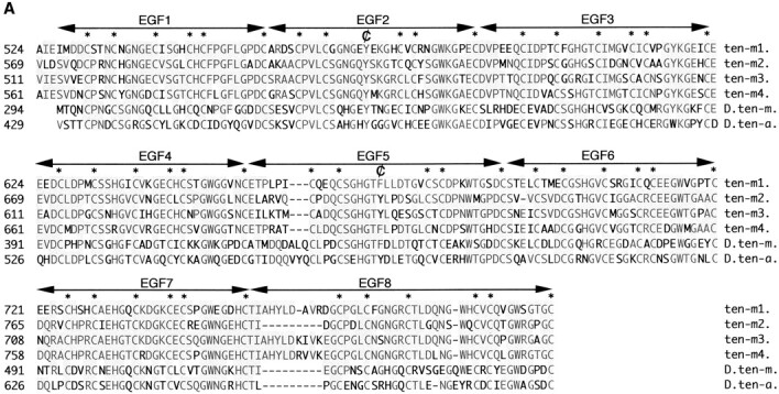
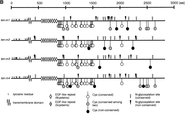
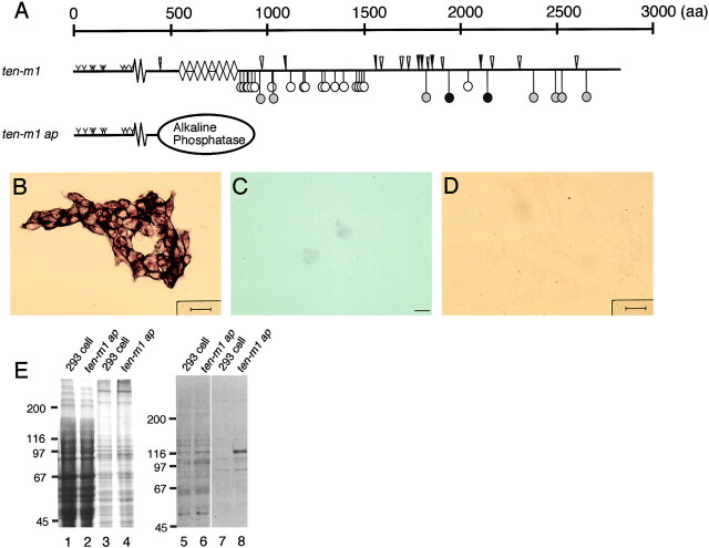
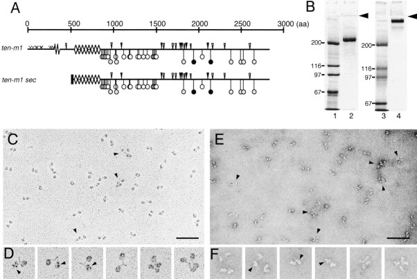
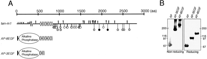
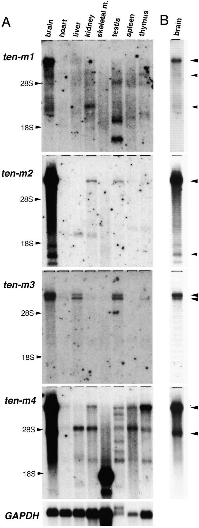
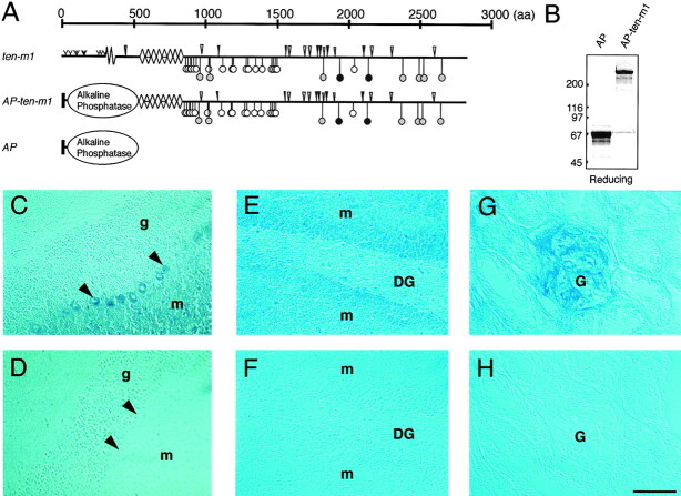
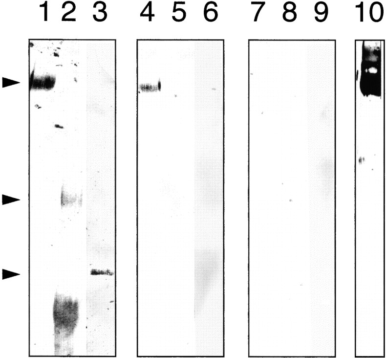
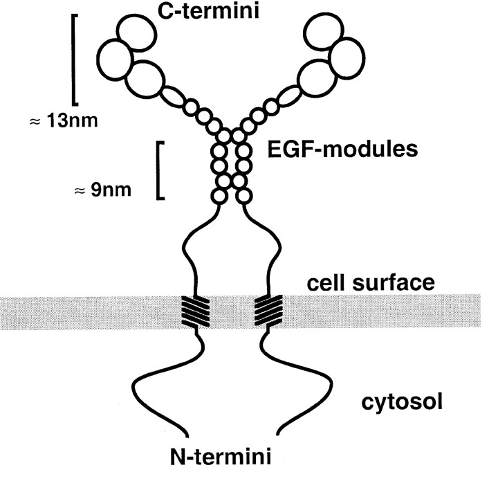
The Drosophila gene ten-m/odz is the only pair rule gene identified to date which is not a transcription factor. In an attempt to analyze the structure and the function of ten-m/odz in mouse, we isolated four murine ten-m cDNAs which code for proteins of 2,700-2, 800 amino acids. All four proteins (Ten-m1-4) lack signal peptides at the NH2 terminus, but contain a short hydrophobic domain characteristic of transmembrane proteins, 300-400 amino acids after the NH2 terminus. About 200 amino acids COOH-terminal to this hydrophobic region are eight consecutive EGF-like domains. Cell transfection, biochemical, and electronmicroscopic studies suggest that Ten-m1 is a dimeric type II transmembrane protein. Expression of fusion proteins composed of the NH2-terminal and hydrophobic domain of ten-m1 attached to the alkaline phosphatase reporter gene resulted in membrane-associated staining of the alkaline phosphatase. Electronmicroscopic and electrophoretic analysis of a secreted form of the extracellular domain of Ten-m1 showed that Ten-m1 is a disulfide-linked dimer and that the dimerization is mediated by EGF-like modules 2 and 5 which contain an odd number of cysteines. Northern blot and immunohistochemical analyses revealed widespread expression of mouse ten-m genes, with most prominent expression in brain. All four ten-m genes can be expressed in variously spliced mRNA isoforms. The extracellular domain of Ten-m1 fused to an alkaline phosphatase reporter bound to specific regions in many tissues which were partially overlapping with the Ten-m1 immunostaining. Far Western assays and electronmicroscopy demonstrated that Ten-m1 can bind to itself.


Figures

Figure 1
Figure 1
Protein sequence of EGF-like modules and structure of mouse Ten-m 1–4. (A) EGF-like domains of mouse Ten-m 1–4, Drosophila Ten-m, and Drosophila Ten-a. Conserved cysteines are indicated by asterisks. Substitutions of cysteines by aromatic amino acids are indicated by the ¢ symbols. Arrows indicate the size of single EGF modules. (B) Overview of the proposed structural organization of the four mouse ten-m proteins. Tyrosine residues in the cytosolic part and potential N-glycosylation sites and cysteines in the extracellular part are indicated, except the 46 cysteine residues within EGF motifs which are marked in A.
Figure 1
Figure 1
Protein sequence of EGF-like modules and structure of mouse Ten-m 1–4. (A) EGF-like domains of mouse Ten-m 1–4, Drosophila Ten-m, and Drosophila Ten-a. Conserved cysteines are indicated by asterisks. Substitutions of cysteines by aromatic amino acids are indicated by the ¢ symbols. Arrows indicate the size of single EGF modules. (B) Overview of the proposed structural organization of the four mouse ten-m proteins. Tyrosine residues in the cytosolic part and potential N-glycosylation sites and cysteines in the extracellular part are indicated, except the 46 cysteine residues within EGF motifs which are marked in A.
Figure 2
Figure 2
Expression of the NH2-terminal sequence of Ten-m1 linked to an AP module in HEK 293 cells. (A) Cartoon of Ten-m1 and of the AP fusion protein (ten-m1 ap). Symbols are the same as in Fig. 1 B. (B) Transfected HEK 293 cells show AP activity on the surface of the cell membrane. (C) After treating transfected cells with trypsin the AP activity is markedly reduced. (D) Mock-transfected HEK 293 cells. No AP activity is visible on these cells. (E) Coomassie blue staining (lanes 1–4) and Western blot with anti-CIAP antiserum (lanes 5–8) of proteins which were present in Triton X-114–poor (lanes 1, 2, 5, and 6) and Triton X-114–rich (lanes 3, 4, 7, and 8) phases after partition at 30°C. Experiments were performed in parallel with cell layers of nontransfected 293 cells (lanes 1, 3, 5, and 7) and 293 cells transfected with the AP fusion protein (lanes 2, 4, 6, and 8). Proteins were separated by 7% SDS-PAGE under reducing conditions.
Figure 3
Figure 3
Expression and electronmicroscopic analysis of the extracellular domain of Ten-m1. (A) Cartoon of Ten-m1 and the secreted COOH-terminal part of Ten-m1 (Ten-m1sec). (B) Coomassie blue staining of molecular mass standards (lanes 1 and 3) and purified Ten-m1sec (lanes 2 and 4) separated by 5% SDS-PAGE under reducing (lanes 1 and 2) and nonreducing (lanes 3 and 4) conditions. The arrowhead indicates the beginning of the separating gel. (C–F) Electron micrographs after glycerol spraying/rotary shadowing (C and D) and negative staining (E and F) of Ten-m1sec. Representative fields of molecules (C and E) and selected species of the same material (D and F) are shown. Pairs of spherical domains, connected by thin elongated rods, are visible. Some of the spheres are resolved into three globular subdomains as indicated by arrowheads (D and F). Arrowheads (C and E) also indicate pairs of Ten-m1sec dimers interacting with each other. Bars, 50 nm.
Figure 4
Figure 4
Expression of EGF-like domains derived from Ten-m1 and fused to an AP module. (A) Cartoon of Ten-m1 and the AP fusion proteins. In AP-3EGF the AP module is linked to the first three EGF-like domains and AP-8EGF to the eight EGF-like domains of Ten-m1. Symbols are the same as in Fig. 1 B. (B) Western blot of AP fusion proteins. Supernatants of 293 cells secreting soluble AP alone, AP-3EGF, or AP-8EGF were precipitated with TCA. The precipitated proteins were separated by 6% SDS-PAGE under nonreducing or reducing conditions, blotted, and detected with anti-CIAP antiserum.
Figure 5
Figure 5
Northern blot of tissues using ten-m cDNAs as probes. Poly(A)+ RNA was isolated from the tissues indicated and probed with ten-m1, 2, 3, and 4 cDNAs coding for the cytoplasmic and transmembrane domain and exposed for 4 d (A) or 24 h (B). Arrowheads in B show the alternatively spliced mRNAs present in brain tissue.
Figure 6
Figure 6
Western blot of brain extracts with affinity-purified antibodies against Ten-m1. 3% Triton X-100 extracts of mouse brain (lanes 1 and 2) were precipitated with acetone. Dried precipitates were dissolved in sample buffer, either for 30 min at 70°C (lane 1) or at room temperature (lane 2). Both samples were reduced for 5 min at 95°C before electrophoresis. Lane 3 shows recombinant Ten-m1sec which served as a positive control and size marker. The arrow indicates the position of a second band occasionally occurring in the recombinant Ten-m1sec lane (also faintly visible here), probably reflecting incompletely reduced Ten-m1sec dimers.
Figure 7
Figure 7
Immunolocalization of Ten-m1 in tissue sections. (A and B) Immunostaining of cerebellum. Strong staining is seen in the molecular layer (m) and weaker staining around cells in the granular cell layer (g). Purkinje cells (arrow in B) show no expression of Ten-m1. The boxed area in A is shown at higher magnification in B. (C and D) Immunostaining in the hippocampus. The staining is very strong in the molecular layer (m) of the CA3 region and weaker around neuronal cells in the CA3 region and dentate gyrus. (E and F) Immunostaining in retina and cornea. Strong staining is seen over the photoreceptor inner (IS) and outer (OS) segment. Very faint staining could be detected in the outer plexiform layer (OPL) and no staining could be detected in the outer nuclear layer (ONL) of the retina. (F) The basal cell layer of the corneal epithelium stained strongly for Ten-m1. Linear staining was also observed on the superficial layer of the corneal epithelium. (G) Ten-m1 staining of a lung section. The expression was high in the smooth muscle cell layer of arteries (a), bronchi (b), and veins (v). Low expression could be also observed on alveolar cells. (H) Ten-m1 expression in glomeruli and (I) on spermatides present in the seminiferous tubules of the testes. The signal was absent from spermatogonia, mature sperm (center of the tubules), Sertoli cells, and Leydig cells. Bars, 150 μm in A, C, and D; 30 μm in B, E, F, and H; 60 μm in G and I.
Figure 8
Figure 8
Binding pattern of AP-ten-m1 in tissue sections. (A) Cartoon of Ten-m1, the fusion protein composed of AP-ten-m1 and AP. Symbols are the same as in Fig. 1 B. (B) Coomassie blue staining of the purified AP-ten-m1 and AP separated by 7% SDS-PAGE under reducing conditions. (C–F) Frozen sections derived from cerebellum (C), hippocampus (E), and kidney (G) were incubated with AP-ten-m1. Control sections (D, F, and H) were incubated with AP. Arrowheads indicate Purkinje cells. g, granular layer; m, molecular layer; DG, dentate gyrus; G, glomerulus. Bars, 50 μm in C, D, G, and H; 25 μm in E and F.
Figure 9
Figure 9
Homophilic interaction of Ten-m1. Western blot of the recombinant extracellular domain of Ten-m1 (lanes 1, 4, 7, and 10), recombinant neurocan core protein (lanes 2, 5, and 8), and BSA (lanes 3, 6, and 9), separated on a 5% (Ten-m1 and neurocan) or on a 7% (BSA) SDS-PAGE performed under nonreducing conditions. Lanes 1–3 were stained with amido black. Arrowheads indicate the 400-kD Ten-m1sec dimer, the 250-kD neurocan core protein, and the 67-kD BSA band. Lanes 4–6 were incubated with purified AP-ten-m1 fusion protein, and lanes 7–9 with purified AP alone. Lane 10 was incubated with the purified anti–Ten-m1 antiserum and AP-conjugated secondary antibodies.
Figure 10
Figure 10
Model of mouse Ten-m1. Two Ten-m1 protein chains are inserted into the plasma membrane as a type II transmembrane molecule. EGF-like modules 2 and 5 are engaged in intermolecular bonds. The COOH terminus is divided into three globular domains.

References

    1. Altschul SF, Boguski MS, Gish W, Wootton JC. Issues in searching molecular sequence databases. Nat Genet. 1994;6:119–129. - PubMed
    1. Barr PJ. Mammalian subtilisins: the long-sought dibasic processing endoproteases. Cell. 1991;66:1–3. - PubMed
    1. Baumgartner S, Chiquet-Ehrismann R. Tena, a Drosophilagene related to tenascin, shows selective transcript localization. Mech Dev. 1993;40:165–176. - PubMed
    1. Baumgartner S, Martin D, Hagios C, Chiquet-Ehrismann R. Tenm, a Drosophilagene related to tenascin, is a new pair-rule gene. EMBO (Eur Mol Biol Organ) J. 1994;13:3728–3740. - PMC - PubMed
    1. Binari R, Perrimon N. Stripe-specific regulation of pair-rule genes by hopscotch, a putative Jak family tyrosine kinase in Drosophila. . Genes Dev. 1994;8:300–312. - PubMed

Publication types

MeSH terms