. 1999 May;103(9):1277-85.
doi: 10.1172/JCI6091.

The Ron/STK receptor tyrosine kinase is essential for peri-implantation development in the mouse

Affiliations

The Ron/STK receptor tyrosine kinase is essential for peri-implantation development in the mouse

R S Muraoka et al. J Clin Invest. 1999 May.

Abstract

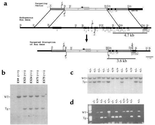
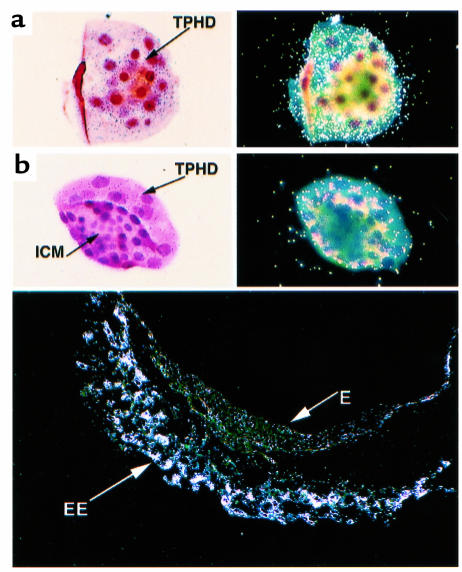
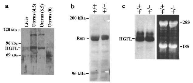
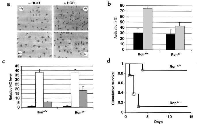
The Ron/STK receptor tyrosine kinase is a member of the c-Met family of receptors and is activated by hepatocyte growth factor-like protein (HGFL). Ron activation results in a variety of cellular responses in vitro, such as activation of macrophages, proliferation, migration, and invasion, suggesting a broad biologic role in vivo. Nevertheless, HGFL-deficient mice grow to adulthood with few appreciable phenotypic abnormalities. We report here that in striking contrast to the loss of its only known ligand, complete loss of Ron leads to early embryonic death. Embryos that are devoid of Ron (Ron-/-) are viable through the blastocyst stage of development but fail to survive past the peri-implantation period. In situ hybridization analysis demonstrates that Ron is expressed in the trophectoderm at embryonic day (E) 3.5 and is maintained in extraembryonic tissue through E7.5, compatible with an essential function at this stage of development. Hemizygous mice (Ron+/-) grow to adulthood; however, these mice are highly susceptible to endotoxic shock and appear to be compromised in their ability to downregulate nitric oxide production. These results demonstrate a novel role for Ron in early mouse development and suggest that Ron plays a limiting role in the inflammatory response.


Figures

Figure 1
Figure 1
Disruption of the mouse Ron gene in embryonic stem (ES) cells and mice. (a) Schematic representation and partial restriction map of the targeting vector (top) used to disrupt the endogenous mouse Ron gene (middle). The predicted organization of the targeted mouse Ron gene following homologous recombination is also shown (bottom). The targeting vector contains sequences homologous to the endogenous Ron gene (gray bars) flanking the HPRT expression cassette (white bar). The HSV-tk gene flanks the targeting vector at the 3′ end (dark gray bar). The exons of the endogenous Ron gene are numbered (1–19, black boxes). The primers used for PCR genotyping are indicated with arrows and are numbered 1–6. The probe used for Southern blot analysis is shown below the endogenous Ron gene. The BamHI fragments of genomic DNA that are complementary to the probe are indicated with the expected sizes (4.7 kb and 3.6 kb). B, BamHI. H, HindIII. S, SalI Sm, SmaI. X, XbaI. (b) Southern blot analysis of BamHI–digested DNA from selection-resistant ES cell clones. The wild-type allele (WT) is represented by a 4.7-kb fragment, while the targeted allele (Tg) is represented by a 3.6-kb fragment. ES9 was included as a wild-type control. (c) Southern blot analysis of BamHI–digested DNA from a single litter generated from a Ron+/– intercrossing. Offspring were genotyped at 7 days postnatal. Of the 12 littermates shown, 8 are Ron+/– (+/–) and 4 are Ron+/+ (+/+). (d) Genotype analysis by PCR of a single litter of E3.5 blastocysts generated from a Ron+/– intercrossing. The wild-type allele is represented by a 523-bp product generated from primers 1 and 2 (see a); the targeted allele is represented by a 274-bp product generated from primers 5 and 6.
Figure 2
Figure 2
In situ hybridization of wild-type embryos at E3.5 and E7.5. The embryos were stained with hematoxylin and eosin and hybridized with a probe complementary to the 3′ end of the mouse Ron transcript. Two distinct orientations of the blastocysts are shown: (a) orientation highlighting the trophectoderm of the blastocysts, and (b) orientation highlighting the inner cell mass. The light-field exposure is shown in the left panels, and the dark-field exposure is shown in the right panels. The bottom panel shows the dark-field exposure of a wild-type embryo isolated with the intact decidua at E7.5. TPHD, trophectoderm. ICM, inner cell mass. EE, extraembryonic tissue. E, embryonic tissue.
Figure 3
Figure 3
Expression of HGFL in the liver and maternal uterus. (a) Expression of HGFL in the pregnant uterus. Uterine tissue was dissected from pregnant wild-type females at 4.5 and 6.5 days postcoitus, or from virgin (0) females. Liver was dissected from virgin females as a positive control for HGFL expression. Proteins were electrophoresed and Western blot analysis was performed. The expected size of HGFL (80 kDa) is indicated, as are the sizes of the molecular weight markers. The identity of the higher-molecular-weight band is unknown, but this band does not correspond to the size expected for HGFL. (b) Expression of Ron in the liver of Ron+/+ and Ron+/– mice. Western blot analysis was performed on protein isolated from the livers of wild-type and Ron+/– mice. The expected size of Ron (∼170 kDa) is indicated, as are the sizes of the molecular weight markers. (c) Expression of HGFL RNA in the livers of Ron+/+ and Ron+/– mice. Total RNA was isolated from the livers of Ron+/+ and Ron+/– mice and analyzed for expression of HGFL mRNA by Northern blot analysis. The position of the mouse HGFL mRNA is indicated, as are the positions of the 18S and 28S ribosomal RNAs. The ethidium bromide–stained agarose gel is shown (at right) to indicate the equivalent loading of samples.
Figure 4
Figure 4
Analysis of macrophage activation, NO synthesis, and septic shock. (a) Resident peritoneal macrophages isolated from Ron+/+ or Ron+/– mice (–HGFL) were treated with 100 ng/mL recombinant human HGFL (+HGFL) and analyzed for morphologic changes, including nuclear condensation, spindle formation, and cytoplasmic elongations. (b) Resident peritoneal macrophages were stimulated with media containing 100 ng/mL recombinant human HGFL (gray bars), or with control media (black bars). Macrophage activation was determined by blinded counting at 1–3 hours following HGFL treatment. Results are presented as the number of activated macrophages per total number of macrophages. Error bars represent the SD (P < 0.009, calculated using the Mann-Whitney test; n = 12 for each genotype; each experiment was repeated 3 times). (c) Resident peritoneal macrophages were stimulated with 1 μg/mL LPS and 100 U/mL IFN-γ in the absence (hatched bars) or presence (gray bars) of 100 ng/mL recombinant human HGFL, or left untreated (black bars). All NO values were calculated in reference to the amount of NO in untreated wild-type macrophages, which was set at a value of 1. The levels of NO in untreated macrophages from Ron+/+ and Ron+/– mice did not differ significantly. All values represent the average values obtained from 3 sets of mice (n = 12 for each genotype). Each experiment was assayed in triplicate. Error bars represent the SD (P < 0.03, calculated using the Mann-Whitney test). (d) Increased susceptibility of Ron+/– mice to LPS-induced septic shock. Ron+/+ or Ron+/– mice were administered 25 μg/g LPS by peritoneal injection and then monitored daily for mortality over a 14-day period. Results are presented as “cumulative survival,” which is the number of surviving mice per total number of mice treated with LPS (n = 8 for each genotype). The 2 groups of mice were compared by Kaplan-Meier survival analysis and were found to be statistically different, with a P value of 0.0011 by the Mantel-Cox test.

References

    1. Bottaro DP, et al. Identification of the hepatocyte growth factor receptor as the c-met proto-oncogene. Science. 1991;251:802–804. - PubMed
    1. Naldini L, et al. Scatter factor and hepatocyte growth factor are indistinguishable ligands for the Met receptor. EMBO J. 1991;10:2867–2878. - PMC - PubMed
    1. Ronsin C, Muscatelli F, Mattei MG, Breathnach R. A novel putative receptor tyrosine kinase of the Met family. Oncogene. 1993;8:1195–1202. - PubMed
    1. Gaudino G, et al. Ron is a heterodimeric tyrosine kinase receptor activated by the HGF homologue MSP. EMBO J. 1994;13:3524–3532. - PMC - PubMed
    1. Schmidt C, et al. Scatter factor/hepatocyte growth factor is essential for liver development. Nature. 1995;373:768–771. - PubMed

Publication types