. 1999 May;103(9):1287-98.
doi: 10.1172/JCI6576.

Analysis of the role of microsomal triglyceride transfer protein in the liver of tissue-specific knockout mice

Affiliations

Analysis of the role of microsomal triglyceride transfer protein in the liver of tissue-specific knockout mice

M Raabe et al. J Clin Invest. 1999 May.

Abstract

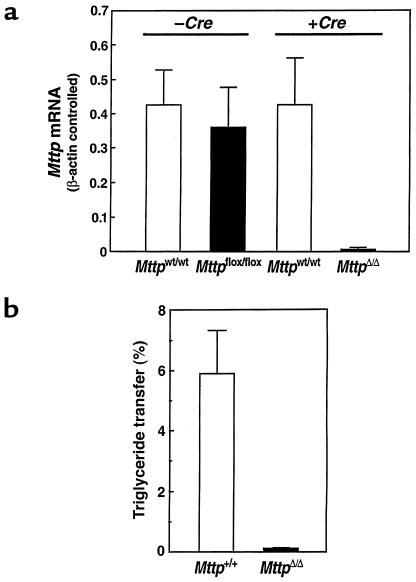
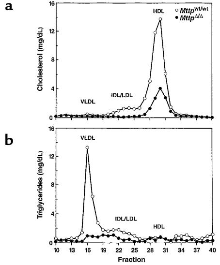
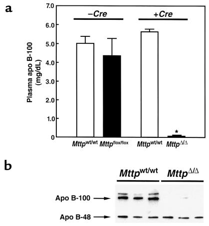
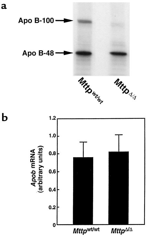
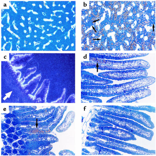
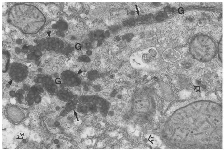
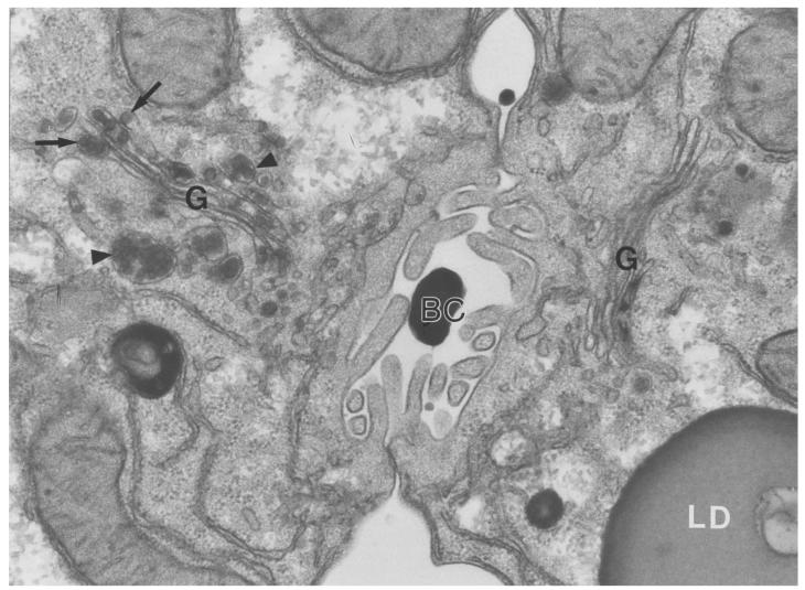
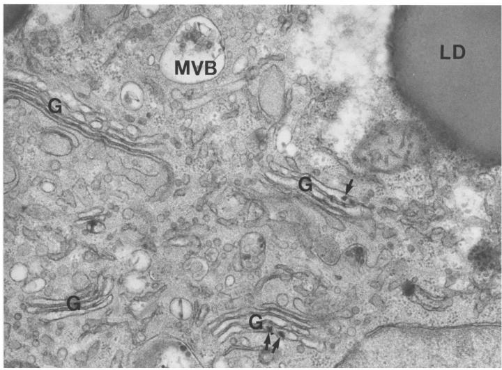
A deficiency in microsomal triglyceride transfer protein (MTP) causes the human lipoprotein deficiency syndrome abetalipoproteinemia. However, the role of MTP in the assembly and secretion of VLDL in the liver is not precisely understood. It is not clear, for instance, whether MTP is required to move the bulk of triglycerides into the lumen of the endoplasmic reticulum (ER) during the assembly of VLDL particles. To define MTP's role in hepatic lipoprotein assembly, we recently knocked out the mouse MTP gene (Mttp). Unfortunately, achieving our objective was thwarted by a lethal embryonic phenotype. In this study, we produced mice harboring a "floxed" Mttp allele and then used Cre-mediated recombination to generate liver-specific Mttp knockout mice. Inactivating the Mttp gene in the liver caused a striking reduction in VLDL triglycerides and large reductions in both VLDL/LDL and HDL cholesterol levels. The Mttp inactivation lowered apo B-100 levels in the plasma by >95% but reduced plasma apo B-48 levels by only approximately 20%. Histologic studies in liver-specific knockout mice revealed moderate hepatic steatosis. Ultrastructural studies of wild-type mouse livers revealed numerous VLDL-sized lipid-staining particles within membrane-bound compartments of the secretory pathway (ER and Golgi apparatus) and few cytosolic lipid droplets. In contrast, VLDL-sized lipid-staining particles were not observed in MTP-deficient hepatocytes, either in the ER or in the Golgi apparatus, and there were numerous cytosolic fat droplets. We conclude that MTP is essential for transferring the bulk of triglycerides into the lumen of the ER for VLDL assembly and is required for the secretion of apo B-100 from the liver.


Figures

Figure 1
Figure 1
Generation and characterization of a floxed Mttp allele. (a) Schematic of the sequence-replacement gene-targeting strategy. The map of the wild-type Mttp allele (Mttpwt) spans the Mttp promoter (the sequences upstream from exon 1) and exons 1–3. The location of the 1.3-kb 3′ flanking probe (5) is indicated. Recombination of the gene-targeting vector with the cognate sequences in the chromosomal DNA produces a mutant Mttp allele, Mttpflox, in which the promoter and exon 1 of the Mttp allele are flanked by loxP sites (filled triangles). An additional loxP site is located downstream from the neo. Cre-mediated excision of both the neo and the promoter/exon 1 fragment produces a null Mttp allele (designated MttpΔ). (b) Illustration of the PvuII fragments produced by different Cre-mediated recombination events: excision of both the neo and the promoter/exon 1 fragment (in an MttpΔ allele), excision of the promoter/exon 1 fragment alone (in an MttpΔex1 allele), and excision of the neo alone (in an MttpΔneo allele). (c) A Southern blot of PvuII-digested genomic DNA from offspring of Mttpwt/flox/deleter-Cre intercrosses. The blot was hybridized with the 3′ flanking probe. Each of the 3 possible Cre-mediated recombination events was observed (MttpΔex1, MttpΔneo, and MttpΔ). Southern blots of PvuII-cleaved genomic DNA did not distinguish between the Mttpflox and MttpΔex1 alleles. The mouse Mttp gene contains SacI sites located 2 kb 5′ and 2.5 kb 3′ to the gene fragment illustrated in a. The gene-targeting event introduced a new SacI site. The recombination events could also be analyzed, therefore, with SacI-cleaved genomic DNA (Mttpwt, 16.5 kb; Mttpflox, 8 kb; MttpΔneo, 7 kb; MttpΔex1, 12.5 kb; and MttpΔ, 11.5 kb). (d) Southern blot illustrating Cre-mediated recombination in different tissues of an Mttpflox/flox/Mx1-Cre mouse in which Cre expression had been induced with pIpC. DNA samples from various tissues were digested with PvuII; the blot was hybridized with the 3′ flanking probe.
Figure 2
Figure 2
Inactivation of the Mttp gene in the liver. (a) Bar graphs illustrating Mttp mRNA levels in groups of Mttpwt/wt and Mttpflox/flox mice (no Cre expression) (n = 5 in each group) and groups of Mttpwt/wt/Mx1-Cre and Mttpflox/flox/Mx1-Cre mice (n = 5 in each group) where Cre expression had been induced with pIpC. Mttp mRNA levels were determined with β-actin–controlled RPAs. (b) A bar graph illustrating MTP activity levels in livers from groups (n = 5) of Mttpwt/wt and Mttpflox/flox mice that had been treated with Cre adenovirus. Each bar graph shows mean ± SD.
Figure 3
Figure 3
Distribution of cholesterol (a) and triglycerides (b) within the plasma lipoproteins of Mttpwt/wt and MttpΔ/Δ mice. Plasma (200 μL) was pooled from 5 females of each genotype after a 4-hour fast and fractionated on an FPLC column (36, 37). The MttpΔ/Δ mice were produced by treating Mttpflox/flox mice with Cre adenovirus. In the 2 experiments, the sum of the cholesterol levels in the HDL fractions of MttpΔ/Δ mice were reduced by 43% and 50% compared with those in Mttpwt/wt mice.
Figure 4
Figure 4
Plasma apo B levels in Mttpwt/wt and MttpΔ/Δ mice. (a) Bar graphs illustrating apo B-100 concentrations (means ± SD) in the plasma of Mttpwt/wt/Mx1-Cre and Mttpflox/flox/Mx1-Cre mice, both before and after pIpC induction of Cre expression. Apo B-100 concentrations were assessed with an mAb-based RIA. (b) Analysis of plasma apo B levels in MttpΔ/Δ mice (Mttpflox/flox/Mx1-Cre mice treated with pIpC), as judged by Western blots of SDS-polyacrylamide gels. Plasma samples (obtained after a 4-hour fast) were size-fractionated on a 4% SDS-polyacrylamide gel, and Western blots were performed with a rabbit antiserum specific for mouse apo B (35).
Figure 5
Figure 5
Apo B secretion and Apob mRNA levels in MttpΔ/Δ mice. (a) Apo B accumulation in the medium from Mttpwt/wt and MttpΔ/Δ primary hepatocytes. The MttpΔ/Δ mice were produced by treating Mttpflox/flox mice with Cre adenovirus. Primary hepatocytes were grown in the presence of [35S]methionine/cysteine; the apo B proteins were then immunoprecipitated and resolved on SDS-polyacrylamide gels. Large amounts of apo B-48 secretion from MttpΔ/Δ primary hepatocytes were observed in this experiment and in 2 subsequent experiments. (b) A bar graph illustrating Apob mRNA levels in the livers of Mttpwt/wt and MttpΔ/Δ mice (n = 10 in each group; P = 0.54). The MttpΔ/Δ mice were produced by treating Mttpflox/flox/Mx1-Cre mice with pIpC. The bar graph shows mean ± SD.
Figure 6
Figure 6
Liver and intestinal histology in Mttpwt/wt and MttpΔ/Δ mice. (a) Osmium tetroxide–stained section of the liver of an Mttpwt/wt mouse. (b) Osmium tetroxide–stained section of the liver of an MttpΔ/Δ mouse (produced by treating Mttpflox/flox/Mx1-Cre mice with pIpC). The yellow staining represents intracellular fat droplets. Arrows point to rare hepatocytes that do not appear to have a significant amount of cytosolic fat droplets. (c) In situ hybridization of the duodenum from a wild-type mouse, demonstrating MTP expression in the villus enterocytes. The location of the crypt cells are denoted by a white arrow. (d and e) Osmium tetroxide–stained section of the duodenum from 2 different MttpΔ/Δ mice). Discrete patches of lipid-filled villus enterocytes (arrows) were observed and were confined to the lower half of the villus. The MTP activity level in the intestine was ∼60% lower in pIpC-treated Mttpflox/flox/Mx1-Cre mice than in wild-type mice, suggesting that a large percentage of the intestinal epithelial cells probably had an MttpΔ/flox genotype. (f) Osmium tetroxide–stained section of the duodenum from an Mttpwt/wt mouse.
Figure 7
Figure 7
Electron micrograph of the Golgi apparatus region of an Mttpwt/wt hepatocyte, illustrating nascent VLDL particles in the smooth ER (open arrows), in the Golgi stacks (arrows), and in grapelike clusters within forming Golgi apparatus secretory vesicles (arrowheads). The majority of the particles in this image are 800–1,400 Å. We examined more than 50 electron micrographs of Mttpwt/wt hepatocytes; VLDL-sized particles were observed in the ER and Golgi of each and every hepatocyte. Some of the Mttpwt/wt hepatocytes had smaller VLDL particles (400–800 Å) than illustrated in this figure. ×36,000.
Figure 8
Figure 8
Electron micrograph of 2 adjoining hepatocytes from an MttpΔ/Δ mouse (a pIpC-treated Mttpflox/flox/Mx1-Cre mouse), providing an example of the rare ultrastructural heterogeneity in hepatocytes from these mice. The cell to the right of the bile canaliculus (BC), almost certainly an MttpΔ/Δ hepatocyte, contained numerous cytosolic lipid droplets (LD) and a complete absence of VLDL-sized, lipid-staining particles within the Golgi apparatus (G). The cell on the right of the BC was typical of >98% of hepatocytes in MttpΔ/Δ mice. The cell to the left of the BC, likely a MttpΔ/flox hepatocyte, had few or no cytosolic lipid droplets and small (300–550 Å) VLDL in Golgi (G) stacks (arrows) and secretory vesicles (arrowheads). We occasionally observed some irregularly shaped lipid-staining “smudges” in the Golgi stacks of MttpΔ/Δ hepatocytes; the nature of this particulate matter is not known. Less than 2% of hepatocytes in MttpΔ/Δ mice were similar to the cell on the left. These rare lipoprotein-producing cells almost certainly had not been converted tMttpΔ/Δ genotype. ×36,000.
Figure 9
Figure 9
Electron micrograph of a hepatocyte from the liver of an MttpΔ/Δ mouse, illustrating the ultrastructural characteristics of the vast majority of hepatocytes. At the ultrastructural level, the most striking feature of the MttpΔ/Δ hepatocytes was the complete absence of VLDL-sized, lipid-staining particles within either the ER or the Golgi apparatus (G). Four Golgi complexes are imaged in this electron micrograph. The MttpΔ/Δ hepatocytes contained numerous cytosolic lipid droplets (LD). Small and irregularly shaped lipid-staining material was occasionally observed within the Golgi cisternae (arrows). The smooth ER is widely dispersed in hepatocytes. We scrutinized more than 100 electron micrographs of MttpΔ/Δ hepatocytes and did not observe round, VLDL-sized, lipid-staining particles in the ER. ×36,000.

References

    1. Gordon DA. Recent advances in elucidating the role of the microsomal triglyceride transfer protein in apolipoprotein B lipoprotein assembly. Curr Opin Lipidol. 1997;8:131–137. - PubMed
    1. Wetterau JR, et al. An MTP inhibitor that normalizes atherogenic lipoprotein levels in WHHL rabbits. Science. 1998;282:751–754. - PubMed
    1. Nielsen LB, et al. Genes for apolipoprotein B and microsomal triglyceride transfer protein are expressed in the heart. Evidence that the heart has the capacity to synthesize and secrete lipoproteins. Circulation. 1998;98:13–16. - PubMed
    1. Borén J, Véniant MM, Young SG. Apo B100–containing lipoproteins are secreted by the heart. J Clin Invest. 1998;101:1197–1202. - PMC - PubMed
    1. Raabe M, et al. Knockout of the abetalipoproteinemia gene in mice: reduced lipoprotein secretion in heterozygotes and embryonic lethality in homozygotes. Proc Natl Acad Sci USA. 1998;95:8686–8691. - PMC - PubMed

Publication types