. 1999 May;103(9):1299-307.
doi: 10.1172/JCI4586.

Tissue inhibitor of metalloproteinase-2 stimulates mesenchymal growth and regulates epithelial branching during morphogenesis of the rat metanephros

Affiliations

Tissue inhibitor of metalloproteinase-2 stimulates mesenchymal growth and regulates epithelial branching during morphogenesis of the rat metanephros

J Barasch et al. J Clin Invest. 1999 May.

Abstract

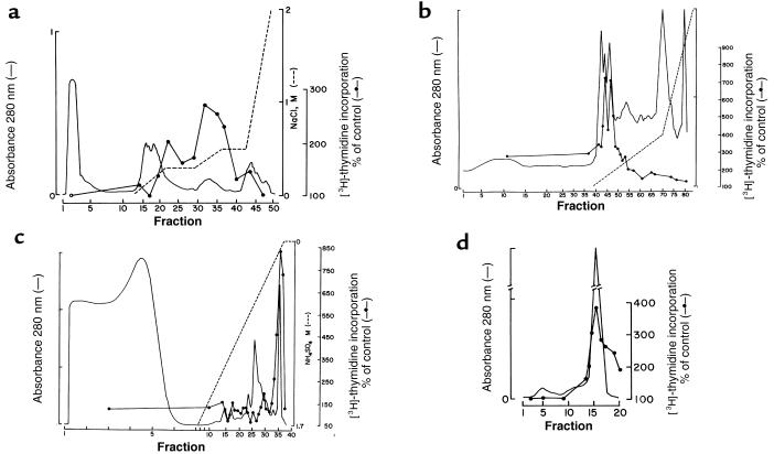
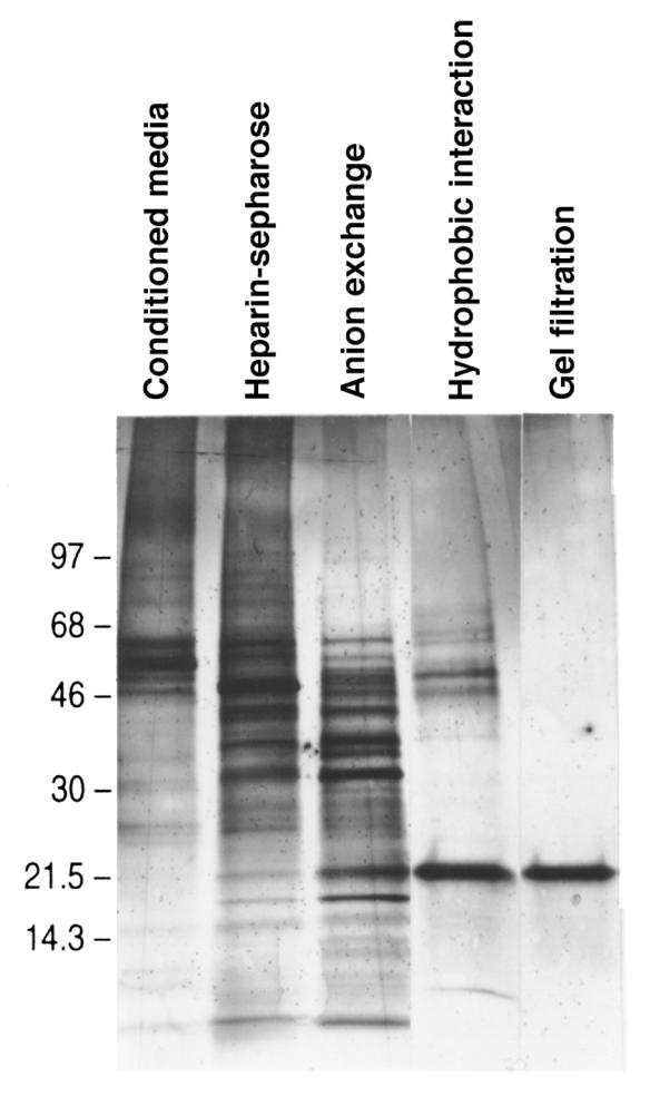
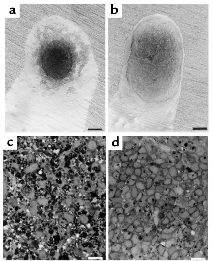
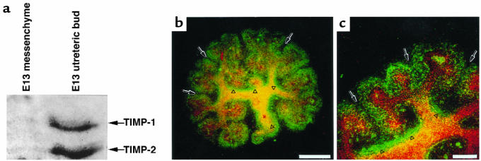
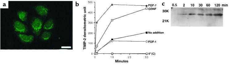
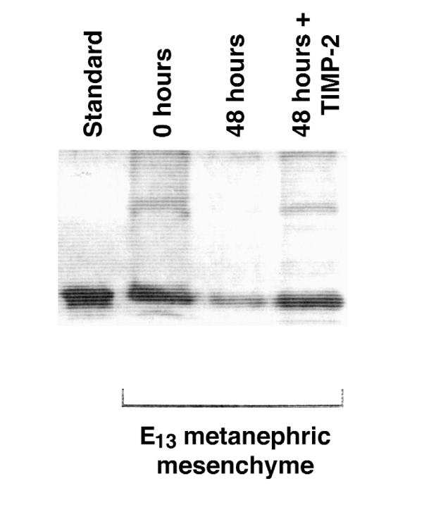
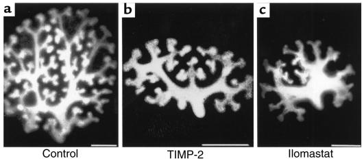
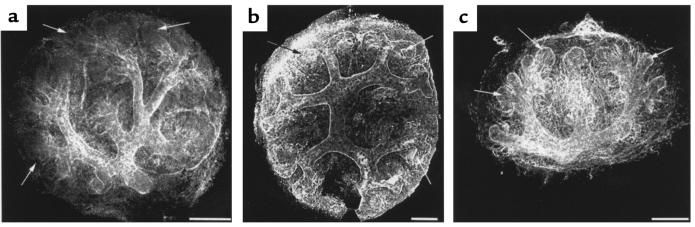
Development of the embryonic kidney results from reciprocal signaling between the ureteric bud and the metanephric mesenchyme. To identify the signaling molecules, we developed an assay in which metanephric mesenchymes are rescued from apoptosis by factors secreted from ureteric bud cells (UB cells). Purification and sequencing of one such factor identified the tissue inhibitor of metalloproteinase-2 (TIMP-2) as a metanephric mesenchymal growth factor. Growth activity was unlikely due to TIMP-2 inhibition of matrix metalloproteinases because ilomastat, a synthetic inhibitor of these enzymes, had no mesenchymal growth action. TIMP-2 was also involved in morphogenesis of the ureteric bud, inhibiting its branching and changing the deposition of its basement membrane; these effects were due to TIMP-2 inhibition of matrix metalloproteinases, as they were reproduced by ilomastat. Thus, TIMP-2 regulates kidney development by at least 2 distinct mechanisms. In addition, TIMP-2 was secreted from UB cells by mesenchymal factors that are essential for ureteric bud development. Hence, the mesenchyme synchronizes its own growth with ureteric morphogenesis by stimulating the secretion of TIMP-2 from the ureteric bud.


Figures

Figure 1
Figure 1
Isolation of TIMP, a metanephric mesenchymal growth factor, from medium conditioned by UB cells. Medium conditioned by UB cells was fractionated by heparin-Sepharose chromatography (a). Three peaks of activity were found (100 μg of each fraction was assayed), and fractions 30–40 were pooled and further purified by a Mono Q anion exchanger (b). Active fractions (fractions 42–48; 10 μg of each fraction assayed) were pooled and further purified by phenyl-Sepharose hydrophobic interaction chromatography (c). Activity copurified with a single protein that we purified to homogeneity by Superdex-75 gel filtration (d). In c and d, 10% volume of each fraction was assayed for [3H]thymidine incorporation.
Figure 2
Figure 2
Isolation of TIMP-2. Silver stain of SDS-PAGE gel of medium conditioned by UB cells, and active fractions from the heparin-Sepharose, anion exchange (2 μg each), hydrophobic, and gel filtration chromatographies (5% volume each).
Figure 3
Figure 3
Rescue of isolated E13 metanephric mesenchymes from apoptosis by TIMP-2. Metanephric mesenchyme involutes (a) and is replaced by apoptotic bodies (c) when cultured for 48 hours in tissue culture medium (MEM with 10% FCS). In contrast, the addition of rTIMP-2 (2 μg/mL) rescues metanephric mesenchymal cells from apoptosis (b). Compared with mesenchymes incubated in basal medium, few apoptotic bodies are found when rTIMP-2 is included (d). Scale bars in a and b: 125 μm. Scale bars in c and d: 10 μm.
Figure 4
Figure 4
Location of TIMP-2 in embryonic kidney. (a) Reverse zymography of isolated E13 mesenchymes and ureteric buds showing prominent localization of TIMP-2 antiprotease activity in the ureteric compartment. (b and c) Confocal microscopy of TIMP-2 immunofluorescence; a three-dimensional X-Y projection of serial cuts through the kidney. TIMP-2 immunoreactivity (green = fluorescein) is found in the condensed metanephric mesenchyme but not in mesenchymal cells that are more distant from the ureteric bud (arrows). Particularly prominent staining is found in the basement matrix surrounding branches of the ureteric bud (arrowheads), which is defined by binding of D. biflorus, a lectin specific for the ureteric bud (red = rhodamine). Scale bar in b: 140 μm. Scale bar in c: 100 μm.
Figure 5
Figure 5
Secretion of TIMP-2 from UB cells is stimulated by mesenchymal proteins that regulate growth and branching of the ureteric bud. (a) UB cells contain immunoreactive TIMP-2. (b) Constitutive secretion of TIMP-2 from UB cells in culture. UB cells were incubated in serum-free MEM, and aliquots collected at the indicated times were assayed by reverse zymography for TIMP-2. (c) Secretion of TIMP-2 is stimulated by GDNF and FGF-7 (100 ng/mL; P < 0.02) but not by FGF-1 (100 ng/mL). At 4°C, both basal and stimulated secretion of TIMP-2 are abolished. Quantification of TIMP-2 secretion was derived from laser densitometry of reverse zymograms. The curves are averaged from 4 independent experiments and analyzed by ANOVA. Scale bar in a: 100 μm.
Figure 6
Figure 6
GDNF in metanephric mesenchymal cells that were rescued from apoptosis by incubation with TIMP-2. GDNF was detected by immunoblots in 6 freshly isolated E13 mesenchymes (0 hours) and in 6 mesenchymes maintained in culture by rTIMP-2 for 48 hours (48 hours + TIMP-2). In contrast, little reactivity remains in 6 untreated mesenchymes (48 hours). Standard is 25 ng of recombinant rat GDNF.
Figure 7
Figure 7
TIMP-2 inhibits ureteric branching in vitro. E13 kidneys were cultured for 4 days with rTIMP-2 (2 μg/mL), and the ureteric bud was then viewed with D. biflorus lectin. Treatment with rTIMP-2 inhibits branching of the ureteric bud (b), as compared with kidneys cultured in basal medium (a). Ilomastat (2 μM), an inhibitor of matrix metalloproteinases, also inhibited branching of the ureteric bud. Images are three-dimensional X-Y projections of serial confocal cuts through the kidney. Scale bars: 300 μm.
Figure 8
Figure 8
TIMP-2 enhances matrix deposition. E13 kidneys were cultured for 4 days with rTIMP-2 (2 μg/mL) and stained with antibodies to collagen IV. The entire ureteric tree is stained by collagen IV antibodies after rTIMP-2 treatment (b), including tip regions (arrows), while only proximal parts of the ureteric bud are obviously ensheathed by collagen IV in control kidneys (a). A few tips of the ureteric bud are indicated (arrow). (c) Similar to the effect of rTIMP-2, treatment of E13 kidneys with ilomastat (2 μM) resulted in matrix deposition throughout the ureteric tree. X-Y projection of serial cuts through the kidney. Scale bars: 100 μm.
Figure 9
Figure 9
TIMP-2 localizes to the basement membrane of the ureteric bud. (a) Immuno-EM of E14 kidneys showing the localization of TIMP-2 to basement membranes of a cleft between 2 branches of the ureteric tree. (b) In contrast, little TIMP-2 is found at the tips of the ureteric bud, an area devoid of basement membrane. Note the curvature of the basal surface of the epithelia that define the tip of the ureteric bud. M, mesenchyme. UB, ureteric bud. Scale bars: 200 nm.

References

    1. Kreidberg JA, et al. WT-1 is required for early kidney development. Cell. 1993;74:679–691. - PubMed
    1. Sanchez M, et al. Renal agenesis and the absence of enteric neurons in mice lacking GDNF. Nature. 1996;382:70–73. - PubMed
    1. Pichel J, et al. Defects in enteric innervation and kidney development in mice lacking GDNF. Nature. 1996;382:73–76. - PubMed
    1. Moore M, et al. Renal and neuronal abnormalities in mice lacking GDNF. Nature. 1996;382:76–79. - PubMed
    1. Schuchardt A, et al. Defects in the kidney and the enteric nervous system of mice lacking the tyrosine kinase receptor ret. Nature. 1994;367:380–383. - PubMed

Publication types

MeSH terms