. 1999 May 15;517 ( Pt 1)(Pt 1):121-34.
doi: 10.1111/j.1469-7793.1999.0121z.x.

A non-capacitative pathway activated by arachidonic acid is the major Ca2+ entry mechanism in rat A7r5 smooth muscle cells stimulated with low concentrations of vasopressin

Affiliations

A non-capacitative pathway activated by arachidonic acid is the major Ca2+ entry mechanism in rat A7r5 smooth muscle cells stimulated with low concentrations of vasopressin

L M Broad et al. J Physiol. .

Abstract

1. Depletion of the Ca2+ stores of A7r5 cells stimulated Ca2+, though not Sr2+, entry. Vasopressin (AVP) or platelet-derived growth factor (PDGF) stimulated Sr2+ entry. The cells therefore express a capacitative pathway activated by empty stores and a non-capacitative pathway stimulated by receptors; only the former is permeable to Mn2+ and only the latter to Sr2+. 2. Neither empty stores nor inositol 1,4,5-trisphosphate (InsP3) binding to its receptors are required for activation of the non-capacitative pathway, because microinjection of cells with heparin prevented PDGF-evoked Ca2+ mobilization but not Sr2+ entry. 3. Low concentrations of Gd3+ irreversibly blocked capacitative Ca2+ entry without affecting AVP-evoked Sr2+ entry. After inhibition of the capacitative pathway with Gd3+, AVP evoked a substantial increase in cytosolic [Ca2+], confirming that the non-capacitative pathway can evoke a significant increase in cytosolic [Ca2+]. 4. Arachidonic acid mimicked the effect of AVP on Sr2+ entry without stimulating Mn2+ entry; the Sr2+ entry was inhibited by 100 microM Gd3+, but not by 1 microM Gd3+ which completely inhibited capacitative Ca2+ entry. The effects of arachidonic acid did not require its metabolism. 5. AVP-evoked Sr2+ entry was unaffected by isotetrandrine, an inhibitor of G protein-coupled phospholipase A2. U73122, an inhibitor of phosphoinositidase C, inhibited AVP-evoked formation of inositol phosphates and Sr2+ entry. The effects of phorbol esters and Ro31-8220 (a protein kinase C inhibitor) established that protein kinase C did not mediate the effects of AVP on the non-capacitative pathway. An inhibitor of diacylglycerol lipase, RHC-80267, inhibited AVP-evoked Sr2+ entry without affecting capacitative Ca2+ entry or release of Ca2+ stores. 6. Selective inhibition of capacitative Ca2+ entry with Gd3+ revealed that the non-capacitative pathway is the major route for the Ca2+ entry evoked by low AVP concentrations. 7. We conclude that in A7r5 cells, the Ca2+ entry evoked by low concentrations of AVP is mediated largely by a non-capacitative pathway directly regulated by arachidonic acid produced by the sequential activities of phosphoinositidase C and diacylglycerol lipase.


Figures
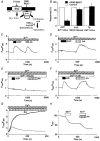
Figure 6
Figure 6. Arachidonic acid mediates the effects of AVP on the non-capacitative pathway
A, targets of the inhibitors used are shown by hammerheads (⊊); pm, plasma membrane. B, inhibition of DAG lipase by RHC-80267 (50 μM, 15 min) selectively inhibited AVP (100 nM)-evoked Sr2+ entry without significantly affecting either AVP-evoked release of Ca2+ from intracellular stores or the capacitative Ca2+ entry evoked by stores that had been emptied by thapsigargin (Tg) (means ±s.e.m., n = 3). C and D, effects of AVP (100 nM) and arachidonic acid (AA, 50 μM) on Sr2+ entry to single A7r5 cells that had been pretreated with ionomycin and thapsigargin are shown for normal cells (C) and for the cell line in which AVP fails to stimulate Sr2+ entry (D). E and F, the results show that both AVP and arachidonic acid stimulate Sr2+ entry which is unaffected by 1 μM Gd3+, but fully inhibited by 100 μM Gd3+. G, in sub-confluent cells, continuous perfusion with arachidonic acid (50 μM) stimulated a sustained Ca2+ entry in almost all cells (thick line; average response from 100 cells), but even 5 μM arachidonic acid (thin line, average response from 5 cells) stimulated a more modest Ca2+ entry in 5 ± 1 % (n = 3 coverslips). The traces are typical of those from 3 independent experiments. H, arachidonic acid (50 μM) did not stimulate Mn2+ influx, indicating that it affected neither membrane integrity nor the capacitative pathway. Traces C-F and H are the average responses of at least 20 individual cells from a single experiment and are typical of 3 independent experiments.
Figure 10
Figure 10. Capacitative and non-capacitative Ca2+ entry pathways
The targets of the inhibitors (hammerheads, ⊊) and the proposed means (arrows) whereby receptors stimulate the two Ca2+ entry pathways are shown.
Figure 1
Figure 1. Capacitative and non-capacitative pathways in A7r5 cells
A and B, cells were treated with thapsigargin (1 μM, Tg) or thapsigargin and ionomycin (1 μM, I/Tg) in Ca2+-free HBS to empty their intracellular stores before addition of either Sr2+ or Ca2+ (1.5 mM) and then AVP (100 nM). A, thapsigargin evoked a transient increase in [Ca2+]i as the intracellular Ca2+ stores released their entire Ca2+ content. The empty stores failed to stimulate Sr2+ entry, but stimulated a substantial entry of Ca2+ through the capacitative pathway. Addition of AVP then caused a decrease in [Ca2+]i due to stimulation of Ca2+ extrusion. B, while pretreatment with ionomycin and thapsigargin to empty the Ca2+ stores failed to stimulate Sr2+ entry, addition of AVP (100 nM) stimulated Sr2+ entry. The same pretreatment was used in all subsequent experiments to empty intracellular Ca2+ stores. C and D, experiments identical to those describd for A and B were performed on the subclone of cells in which the effects of AVP on Sr2+ entry were selectively lost. During many subsequent passages, the cells retained this phenotype and never recovered an ability to respond to AVP with Sr2+ entry. Traces are each representative of at least 10 recordings. In each of the traces recording Sr2+ entry (A-D and Fig. 3), the fluorescence ratios have not been calibrated to [Sr2+]i because although the increase in fluorescence ratio is entirely due to Sr2+ entry (see Fig. 2), the initial fluorescence results from Ca2+ bound to fura-2. It should, however, be noted that fura-2 has a much lower affinity for Sr2+ (Kd= 7.6 μM) than for Ca2+ (Kd= 227 nM) (Byron & Taylor, 1995).
Figure 3
Figure 3. The effect of a low concentration of AVP on Sr2+ entry is more sustained than that of a maximal concentration
A, the Sr2+ entry evoked by AVP (100 nM) is transient, but subsequent addition of PDGF (10 nM) evokes further Sr2+ entry. B, the Sr2+ entry evoked by a submaximal concentration of AVP (1 nM, i) is more sustained than that evoked by a maximal concentration (100 nM, ii).
Figure 2
Figure 2. The fura-2 fluorescence changes evoked by AVP reflect an increase in [Sr2+]i
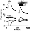
In an experiment similar to that shown in Fig. 1B, fura-2 fluorescence was simultaneously recorded at four excitation wavelengths: 340 and 380 nm to provide the fluorescence ratio (panel i; F340/380) used in all experiments, and at the isoemissive wavelengths for Ca2+ (358 nm) and Sr2+ (363 nm) (panel ii). The results show that the fluorescence changes that follow stimulation of cells with AVP in the presence of extracellular Sr2+ result wholly from an increase in [Sr2+]i.
Figure 9
Figure 9. Low concentrations of AVP preferentially activate non-capacitative Ca2+ entry
A, each trace shows the average change in [Ca2+]i recorded from 4 coverslips of cells stimulated with 1 μM (i), 1 nM (ii), or 100 pM (iii) AVP in the presence (red and black) or absence (blue) of extracellular Ca2+. Blue traces are the responses that result solely from release of intracellular Ca2+ stores. Red traces show responses from cells that were not treated with Gd3+ and therefore represent the normal responses to AVP. Cells shown by the black traces were pretreated with 1 μM Gd3+ for 600 s and therefore represent responses without capacitative Ca2+ entry. B, the Ca2+ signal resulting from Ca2+ mobilization has been subtracted to reveal the total Ca2+ entry component (red) and the Ca2+ entry that persists after Gd3+ treatment (black); the latter therefore represents non-capacitative Ca2+ entry. C, the fraction of the Ca2+ entry mediated by the non-capacitative (Gd3+-resistant; black traces) and capacitative (total Ca2+ entry less that remaining after Gd3+ treatment; red traces) pathways was determined as described in the text. D, the relative contributions of the two pathways 600 s after AVP addition are summarized and show that at low concentrations of AVP, the non-capacitative pathway is the major route for Ca2+ entry (means ±s.e.m., n = 3). E, in the subclone of cells lacking AVP-evoked Sr2+ entry, all Ca2+ entry evoked by 1 nM AVP is blocked by 1 μM Gd3+, indicating that it occurs solely via the capacitative pathway (lines are coded as in A). F, the relative magnitudes (% maximal response) of the Ca2+ mobilization and capacitative Ca2+ entry evoked by three concentrations of AVP suggest that store depletion is tightly coupled to stimulation of the capacitative pathway.
Figure 4
Figure 4. Selective block of the capacitative pathway by Gd3+
A and B, methods similar to those of Fig. 1 were used to examine the effects of Gd3+ (1 μM) on capacitative Ca2+ entry (A) and AVP-evoked Sr2+ entry (B); only the capacitative pathway was blocked by 1 μM Gd3+. C, the concentration-dependent effects of Gd3+ on capacitative Ca2+ entry (○) and AVP-induced Sr2+ entry (•) (means ±s.e.m., n = 3). D and E, Gd3+ (3 μM) irreversibly inhibited capacitative Ca2+ entry (D), but the inhibition of AVP-evoked Sr2+ entry by 100 μM Gd3+ was reversible (E). F, despite the counteracting effect of the stimulation of Ca2+ extrusion by AVP (i, no Gd3+), AVP stimulated an increase in [Ca2+]i when added to cells in which the capacitative pathway had been fully inhibited by Gd3+ (ii, 1 μM). Each trace is typical of at least 3 experiments.
Figure 5
Figure 5. Phosphoinositidase C is required for AVP to stimulate the non-capacitative pathway
A and B, concentration-dependent effects of U73122 (•) and U73343 (○) on AVP (50 nM)-evoked formation of [3H]inositol phosphates (A) and Sr2+ entry (B). Results are means ±s.e.m. of 3 independent experiments. C, U73122 (10 μM) abolished the Sr2+ entry evoked by AVP (100 nM) in thapsigargin-treated cells without affecting that evoked by arachidonic acid (AA, 50 μM). Results are typical of those from 3 similar experiments.
Figure 7
Figure 7. Arachidonic acid metabolism is not required for it to stimulate the non-capacitative pathway
A, in the presence of NDGA (50 μM, thick trace), the Sr2+ entry evoked by AVP (100 nM) was substantially enhanced relative to control (thin trace). The histograms (means ±s.e.m., n = 3) show the fluorescence signal recorded 500 s after additon of AVP in control cells and cells treated with NDGA. B, aspirin (500 μM, thick trace) had no effect on AVP-evoked Sr2+ entry (control, thin trace). Traces are typical records from 3 independent measurements of cell populations.
Figure 8
Figure 8. Depletion of intracellular Ca2+ stores is not required for activation of the non-capacitative pathway
Cells were microinjected with heparin (10 mg ml−1 in injection pipette; thin trace) and then stimulated with PDGF (2.5 nM) in Ca2+-free HBS. Intracellular heparin abolished the Ca2+ mobilization evoked by PDGF (2.5 nM) without affecting Sr2+ entry. Subsequent addition of ionomycin and thapsigargin (I/Tg) confirmed that whereas PDGF had fully emptied the intracellular Ca2+ stores of the control cells (thick trace), the stores of the cells injected with heparin (thin trace) had retained their Ca2+. The traces show results from individual cells typical of 3 similar experiments.

Comment in

References

    1. Akiba S, Kato E, Sato T, Fujii T. Biscolaurine alkaloids inhibit receptor-mediated phospholipase A2 activation probably through uncoupling of a GTP-binding protein from the enzyme in rat peritoneal mast cells. Biochemical Pharmacology. 1992;44:45–50. 10.1016/0006-2952(92)90036-I. - DOI - PubMed
    1. Alonso-Torre SR, Garciá-Sancho J. Arachidonic acid inhibits capacitative calcium entry in rat thymocytes and human neutrophils. Biochimica et Biophysica Acta. 1997;1328:207–213. - PubMed
    1. Berridge MJ. Inositol trisphosphate and calcium signalling. Nature. 1993;361:315–325. - PubMed
    1. Bleasdale JE, Thakur NR, Gremban RS, Bundy GL, Fitzpatrick FA, Smith RJ, Bunting S. Selective inhibition of receptor-coupled phospholipase C-dependent processes in human platelets and polymorphonuclear neutrophils. Journal of Pharmacology and Experimental Therapeutics. 1990;255:756–768. - PubMed
    1. Byron KL, Taylor CW. Spontaneous Ca2+ spiking in a vascular smooth muscle cell line is independent of the release of intracellular Ca2+ stores. Journal of Biological Chemistry. 1993;268:6945–6952. - PubMed

Publication types

LinkOut - more resources