. 1999 May 15;517 ( Pt 1)(Pt 1):287-300.
doi: 10.1111/j.1469-7793.1999.0287z.x.

The pattern of excitation of human lower limb motoneurones by probable group II muscle afferents

Affiliations

The pattern of excitation of human lower limb motoneurones by probable group II muscle afferents

M Simonetta-Moreau et al. J Physiol. .

Abstract

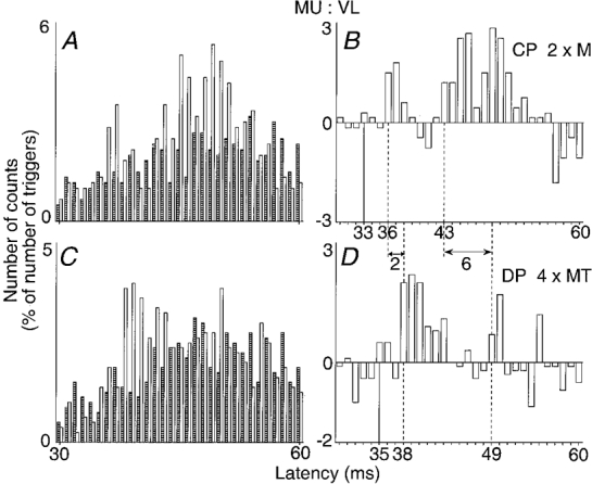
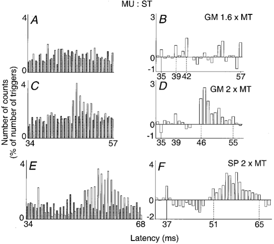
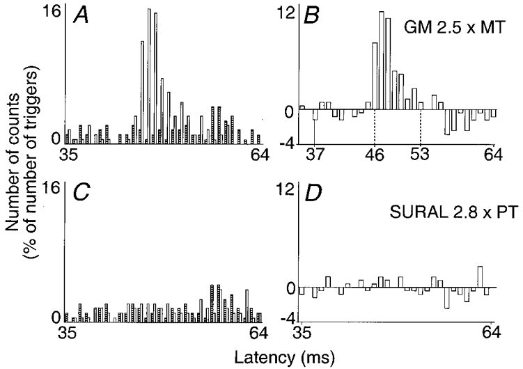
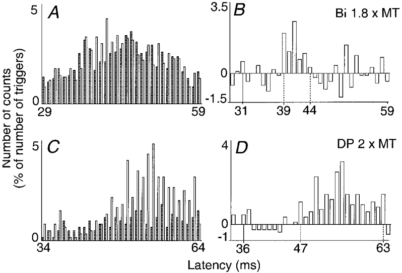
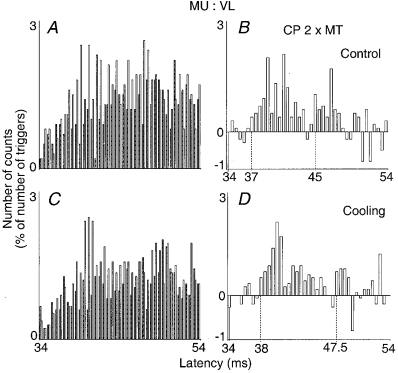
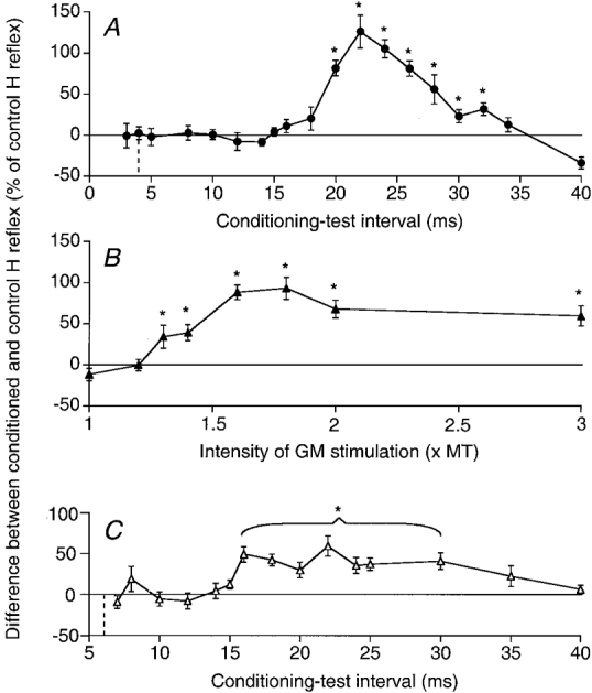
1. Heteronymous group II effects were investigated in the human lower limb. Changes in firing probability of single motor units in quadriceps (Q), biceps (Bi), semitendinosus (ST), gastrocnemius medialis (GM) and tibialis anterior (TA) were studied after electrical stimuli between 1 and 3 times motor threshold (MT) applied to common peroneal (CP), superficial (SP) and deep (DP) peroneal, Bi and GM nerves in those nerve-muscle combinations without recurrent inhibition. 2. Stimulation of the CP and Bi nerves evoked in almost all of the explored Q motor units a biphasic excitation with a low-threshold early peak, attributable to non-monosynaptic group I excitation, and a higher threshold late peak. When the CP nerve was cooled (or the stimulation applied to a distal branch, DP), the increase in latency was greater for the late than for the early peak, indicating that the late excitation is due to stimulation of afferents with a slower conduction velocity than group I fibres, presumably in the group II range. In ST motor units the group II excitation elicited by stimulation of the GM and SP nerves was particularly large and frequent, and the non-monosynaptic group I excitation was often replaced by an inhibition. 3. A late group II-induced excitation from CP to Q motoneurones and from GM and SP to ST motoneurones was also observed when using the H reflex as a test. 4. The electrical threshold and conduction velocity of the largest diameter fibres evoking the group II excitation were estimated to be 2.1 and 0.65 times those of the fastest Ia afferents, respectively. In the combinations tested in the present investigation the group II input seemed to be primarily of muscle origin. 5. The potent heteronymous group II excitation of motoneurones of both flexors and extensors of the knee contrasted with the absence of a group II effect from DP to GM and from GM to TA. In none of the combinations explored was there any evidence for group II inhibition of motoneurones. The possible contribution to postural reactions of the potent group II excitation of thigh motoneurones is discussed.


Figures

Figure 1
Figure 1. Changes in firing probability of one Q motor unit evoked by proximal and distal stimulation of the common peroneal (CP) nerve
A and B, proximal stimulation of the CP nerve (2 × MT) at the upper part of the popliteal fossa. C and D, distal stimulation of the deep peroneal (DP) nerve (4 × MT). The histograms in A and C show discharges of the voluntary activated motor unit in control conditions (hatched columns) and after stimulation of the nerve (open columns). The differences between these two histograms are plotted in B and D. In both cases, the number of counts, expressed as a percentage of the number of the triggers (A and B, 600; C and D, 539) is plotted against the latency from the stimulation (bin width 1 ms). Continuous vertical lines, expected time of arrival of the conditioning Ia volley at the segmental spinal level of the motoneurone (latency of monosynaptic homonymous excitation after stimulation of the femoral nerve, 28 ms; distance between stimulation sites of femoral and CP nerves, 40 cm). Dashed vertical lines, latencies of non-monosynaptic group I and group II excitations. Double-headed horizontal arrows, difference between the latencies of the early peaks (2 ms) and of the late peaks (6 ms) in the two positions of the stimulation.
Figure 4
Figure 4. Changes in firing probability of semitendinosus (ST) motor units after stimulation of the gastrocnemius medialis (GM) and of the superficial peroneal (SP) nerves
A-D, stimulation of the GM nerve at 1.6 (A and B) and 2 (C and D) × MT. E and F, stimulation of the SP nerve at 2 × MT (same subject but another motor unit). Left and right plots and vertical continuous lines as in Fig. 1 (bin width 1 ms). Vertical dotted lines, first and last bins of group I (B) or group II (D and F) excitation. Latency of homonymous monosynaptic excitation from the sciatic nerve, A-D, 31 ms; E and F, 32 ms; distances between stimulations sites of GM and sciatic, and of SP and sciatic nerves, 33 and 38 cm respectively. Number of triggers, A and B, 1105; C and D, 2252; E and F, 1085.
Figure 6
Figure 6. Absence of effect of a sural nerve stimulation
Changes in firing probability of a ST motor unit evoked by stimulation of the GM (A and B, 2.5 × MT) and of the sural (C and D, 2.8 × perception threshold, PT) nerves are compared. Left and right plots and vertical continuous line as in Fig. 1 (bin width 1 ms). Dotted vertical lines as in Fig. 3. Latency of homonymous monosynaptic excitation from the sciatic nerve, 31 ms; distance between the stimulation sites of the sciatic and GM nerves, 35 cm and between those of GM and sural nerves, 38 cm. Number of triggers, A-D, 1099.
Figure 3
Figure 3. Changes in firing probability of a Q (VL) motor unit after stimulation of the biceps (Bi) nerve (A and B, 1.8 × MT) and of a Bi motor unit after stimulation of the DP nerve (C and D, 2 × MT)
Left and right plots and vertical continuous lines as in Fig. 1 (bin width 1 ms). Vertical dotted lines, first and last bins of the group II excitation. A and B, latency of homonymous monosynaptic Ia excitation from the femoral nerve, 29 ms; distance between the sites of stimulation of the femoral and Bi nerves, 20 cm; number of triggers, 1205. C and D, latency of homonymous monosynaptic Ia excitation from the sciatic nerve, 31 ms; distance between the sites of stimulations of DP and sciatic nerves, 35 cm; number of triggers, 599.
Figure 2
Figure 2. Effects of cooling on the changes in firing probability of a Q motor unit evoked by stimulation of the CP nerve (2 × MT)
Results obtained before (A and B) and during (C and D) cooling (20-40 min) of the lower limb are compared (bin width 0.5 ms). Left and right plots and dotted vertical lines as in Fig. 1. Latency of homonymous monosynaptic Ia excitation after femoral nerve stimulation, 27.5 ms; distance between stimulation sites of CP and femoral nerves, 46 cm. Number of triggers: A and B, 1008; C and D, 904. Skin temperature in the popliteal fossa: A and B, 31 °C; C and D, 19 °C.
Figure 5
Figure 5. Facilitation of the ST H reflex elicited by stimulation of the GM and SP nerves
The difference between the amplitude of conditioned and control H reflexes (expressed as a percentage of the control H reflex) is plotted against the conditioning-test interval (A, GM nerve stimulation = 2 × MT, and C, SP stimulation = 2 × MT) or the GM stimulus intensity (B, 22 ms conditioning-test interval). Data from two different subjects (A, B and C). The vertical dotted lines in A and C indicate the ISI corresponding to the simultaneous arrival at the segmental spinal level of the conditioning and test Ia volleys. Each symbol represents the mean of 20 measurements. Vertical bars, 1 standard error of the mean. Asterisks indicate the results which are statistically significant (P < 0.05; note that in C all values between 16 and 30 ms were tested together).

Similar articles

Cited by

References

    1. Burke D, Gracies JM, Mazevet D, Meunier S, Pierrot-Deseilligny E. Non-monosynaptic transmission of the cortical command for voluntary movement in man. The Journal of Physiology. 1994;480:191–207. - PMC - PubMed
    1. Cavallari P, Edgley SA, Jankowska E. Postsynaptic actions of midlumbar interneurones on motoneurones of hind limb muscles in the cat. The Journal of Physiology. 1987;389:675–690. - PMC - PubMed
    1. Chaix Y, Marque Ph, Meunier S, Pierrot-Deseilligny E, Simonetta-Moreau M. Further evidence for non-monosynaptic group I excitation of motoneurones in the human lower limb. Experimental Brain Research. 1997;115:35–46. - PubMed
    1. Corna S, Grasso M, Nardone A, Schieppati M. Selective depression of medium-latency leg and foot muscle responses to stretch by an α2-agonist in humans. The Journal of Physiology. 1995;484:803–809. - PMC - PubMed
    1. Daniel C, Lehmann EL. Henry Scheffé. Annals of Statistics. 1979;7:1149–1161.

Publication types

LinkOut - more resources