. 1999 May;10(5):1297-308.
doi: 10.1091/mbc.10.5.1297.

Different domains of the M-band protein myomesin are involved in myosin binding and M-band targeting

Affiliations
Free PMC article

Different domains of the M-band protein myomesin are involved in myosin binding and M-band targeting

D Auerbach et al. Mol Biol Cell. 1999 May.
Free PMC article

Abstract

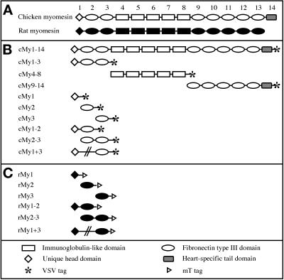
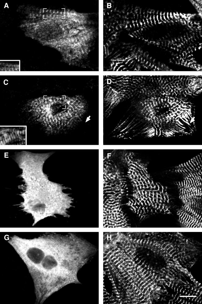
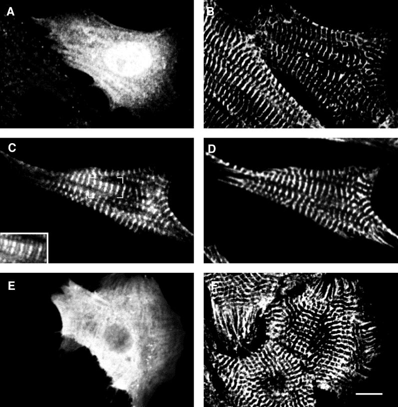
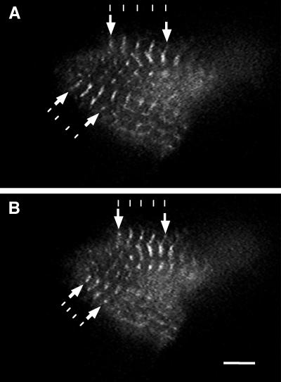
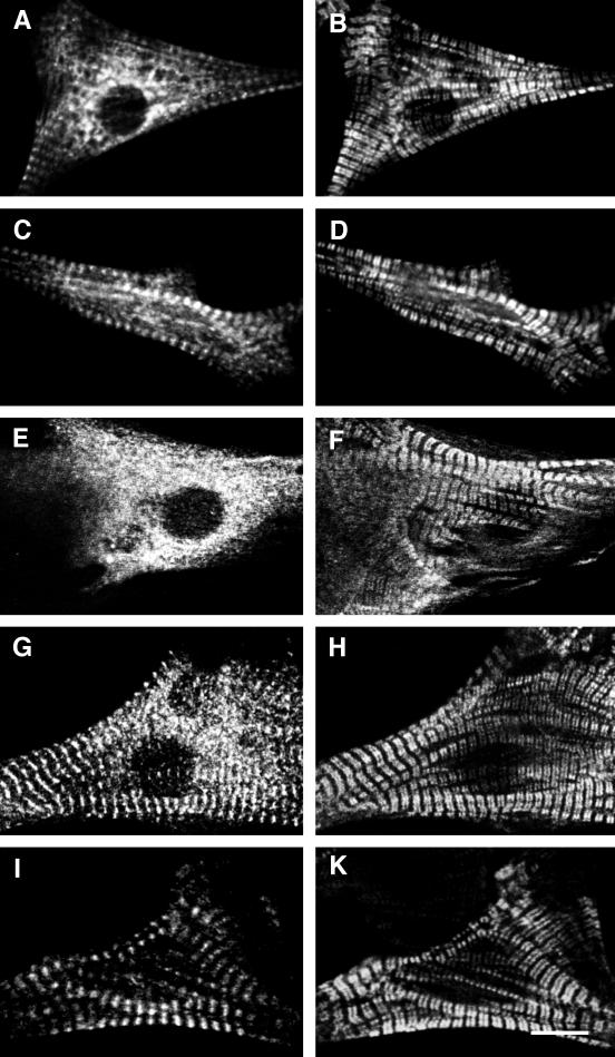
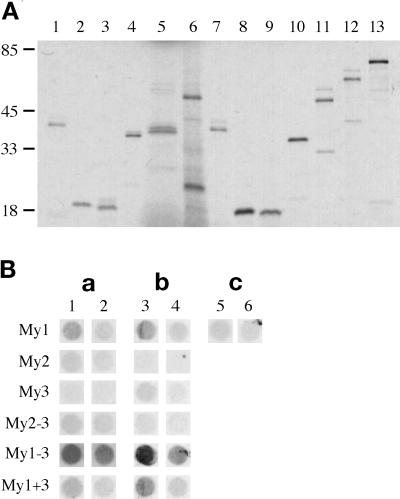
Myomesin is a 185-kDa protein located in the M-band of striated muscle where it interacts with myosin and titin, possibly connecting thick filaments with the third filament system. By using expression of epitope-tagged myomesin fragments in cultured cardiomyocytes and biochemical binding assays, we could demonstrate that the M-band targeting activity and the myosin-binding site are located in different domains of the molecule. An N-terminal immunoglobulin-like domain is sufficient for targeting to the M-band, but solid-phase overlay assays between individual N-terminal domains and the thick filament protein myosin revealed that the unique head domain contains the myosin-binding site. When expressed in cardiomyocytes, the head domains of rat and chicken myomesin showed species-specific differences in their incorporation pattern. The head domain of rat myomesin localized to a central area within the A-band, whereas the head domain of chicken myomesin was diffusely distributed in the cytoplasm. We therefore conclude that the head domain of myomesin binds to myosin but that this affinity is not sufficient for the restriction of the domain to the M-band in vivo. Instead, the neighboring immunoglobulin-like domain is essential for the precise incorporation of myomesin into the M-band, possibly because of interaction with a yet unknown protein of the sarcomere.


Figures

Figure 1
Figure 1
Chicken and rat myomesin truncation and deletion mutants. (A) Domain pattern derived from the cDNAs of chicken and rat myomesin. A head domain with no homology to other known proteins is followed by a conserved arrangement of immunoglobulin-like and fibronectin type III domains. The heart isoform of chicken myomesin contains an additional unique domain at the C terminus. (B) Chicken myomesin truncation and deletion mutants. The VSV epitope was fused C-terminally to all constructs. (C) Rat myomesin truncation and deletion mutants. The mT epitope was fused C-terminally to all constructs. The deletion mutants are depicted by an interrupted line connecting domains one and three in B and C. Abbreviations used in this article to indicate the constructs are shown on the left-hand side.
Figure 2
Figure 2
Full-length chicken myomesin and truncation mutants expressed in NRC. Confocal images of neonatal rat cardiomyocytes expressing full-length chicken heart myomesin cMy1–14 (A and B) and truncation mutants cMy1–3 (C and D), cMy4–8 (E and F), and cMy9–13 (G and H). Cells were stained with an antibody against the VSV epitope (A, C, E, G) and with either an antibody against sarcomeric α-actinin (B, D, H) or an antibody against myomesin (F). Only cMy1–14 and cMy1–3 are incorporated into the M-band region of the myofibrils, as shown by the superimposition of VSV epitope and α-actinin signals in insets A and C. The constructs cMy4–8 and cMy9–14 are diffusely distributed in the cytoplasm. In G, some unspecific interaction of the recombinant protein with the myofibrils can be seen, which probably is an effect of high expression levels. The arrow in D marks NSMFs that incorporate sarcomeric α-actinin but not myomesin. The antibody against myomesin used in F is directed against an epitope in domain 11 and therefore recognizes only endogenous myomesin but not the truncation cMy4–8. Bar, 10 μm.
Figure 3
Figure 3
N-terminal chicken myomesin truncation mutants expressed in NRC. Confocal images of neonatal rat cardiomyocytes expressing constructs cMy1 (A and B), cMy2 (C and D), and cMy3 (E and F). Cells were stained with an antibody against the VSV epitope (A, C, E) and with an antibody against sarcomeric α-actinin (B, D, F). Only the construct cMy2 is targeted to the M-band, whereas cMy1 and cMy3 are diffusely distributed in the cytoplasm of the cells. The inset in C shows a superimposition of the VSV epitope (C) and the α-actinin (D) signals. Bar, 10 μm.
Figure 4
Figure 4
Living cardiomyocyte expressing cMy2–GFP. Single images taken from a video recording of a cardiomyocyte expressing cMy2–GFP showing relaxed (A) and contracted (B) states. cMy2–GFP localizes to the M-bands of the sarcomere and does not inhibit the contractile activity of the cardiomyocyte. The contraction of the sarcomeres can be seen by comparing the decrease in distance between neighboring M-bands in the contracted and uncontracted state (arrows). The lines are drawn to visualize the shortening of the distance between neighboring M-bands. Bar, 5 μm.
Figure 5
Figure 5
N-terminal rat myomesin deletion mutants expressed in NRC. Confocal images of neonatal rat cardiomyocytes expressing constructs rMy1 (A and B), rMy2 (C and D), rMy3 (E and F), rMy2–3 (G and H), and the deletion mutant rMy1 + 3 (I and K). Cells were stained with an antibody against the mT epitope (A, C, E, G, I) and an antibody against MyBP-C (B, D, F, H, K). The constructs rMy1 and rMy2 are targeted to an area within the A-band and to the M-band, respectively, but significant levels of unincorporated protein are present in the cytoplasm. The amount of unincorporated protein is reduced in cells expressing the two-domain constructs rMy2–3 and rMy1 + 3. Bar, 10 μm.
Figure 6
Figure 6
Myosin binding of N-terminal truncation and deletion mutants. (A) SDS-PAGE (8–22%) of chicken and rat constructs expressed in the coupled transcription/translation system. Lanes 1–6, chicken mutants: lane 1, cMy1; lane 2, cMy2; lane 3, cMy3; lane 4, cMy2–3; lane 5, cMy1 + 3; lane 6, cMy1–3. Lanes 7–12, rat mutants: lane 7, rMy1; lane 8, rMy2; lane 9, rMy3; lane 10, rMy2–3; lane 11, rMy1 + 3; lane 12, rMy1–3. Lane 13, luciferase. Molecular weight standards are in kilodaltons. All constructs express proteins of the expected size, although in the case of longer constructs, additional lower bands representing either alternative initiation events or proteolytic degradation products can be observed. (B) Solid-phase binding assay of chicken and rat constructs. The rows are labeled after the domain nomenclature used in Figure 1. a, Chicken constructs; b, rat constructs; c, luciferase control. Different amounts of LMM fragment were used: 1 μg LMM fragment (columns 1, 3, 5) and 100 ng LMM fragment (columns 2, 4, 6).

Similar articles

Cited by

References

    1. Bähler M, Eppenberger HM, Wallimann T. Novel thick filament protein of chicken pectoralis muscle: the 86 kd protein. II. Distribution and localization. J Mol Biol. 1985;186:393–401. - PubMed
    1. Bantle S, Keller S, Haussmann I, Auerbach D, Perriard E, Mühlebach S, Perriard J-C. Tissue-specific isoforms of chicken myomesin are generated by alternative splicing. J Biol Chem. 1996;271:19042–19052. - PubMed
    1. Carlsson E, Thornell LE. Diversification of the myofibrillar M band in rat skeletal muscle during postnatal development. Cell Tissue Res. 1987;248:169–180. - PubMed
    1. Chalfie M, Tu Y, Euskirchen G, Ward WW, Prasher DC. Green fluorescent protein as a marker for gene expression. Science. 1994;263:802–805. - PubMed
    1. Craig R, Offer G. The location of C-protein in rabbit skeletal muscle. Proc R Soc Lond B Biol Sci. 1976;192:325–332. - PubMed

Publication types

MeSH terms

LinkOut - more resources