. 1999 May;10(5):1337-51.
doi: 10.1091/mbc.10.5.1337.

Apg7p/Cvt2p is required for the cytoplasm-to-vacuole targeting, macroautophagy, and peroxisome degradation pathways

Affiliations
Free PMC article

Apg7p/Cvt2p is required for the cytoplasm-to-vacuole targeting, macroautophagy, and peroxisome degradation pathways

J Kim et al. Mol Biol Cell. 1999 May.
Free PMC article

Abstract

Proper functioning of organelles necessitates efficient protein targeting to the appropriate subcellular locations. For example, degradation in the fungal vacuole relies on an array of targeting mechanisms for both resident hydrolases and their substrates. The particular processes that are used vary depending on the available nutrients. Under starvation conditions, macroautophagy is the primary method by which bulk cytosol is sequestered into autophagic vesicles (autophagosomes) destined for this organelle. Molecular genetic, morphological, and biochemical evidence indicates that macroautophagy shares much of the same cellular machinery as a biosynthetic pathway for the delivery of the vacuolar hydrolase, aminopeptidase I, via the cytoplasm-to-vacuole targeting (Cvt) pathway. The machinery required in both pathways includes a novel protein modification system involving the conjugation of two autophagy proteins, Apg12p and Apg5p. The conjugation reaction was demonstrated to be dependent on Apg7p, which shares homology with the E1 family of ubiquitin-activating enzymes. In this study, we demonstrate that Apg7p functions at the sequestration step in the formation of Cvt vesicles and autophagosomes. The subcellular localization of Apg7p fused to green fluorescent protein (GFP) indicates that a subpopulation of Apg7pGFP becomes membrane associated in an Apg12p-dependent manner. Subcellular fractionation experiments also indicate that a portion of the Apg7p pool is pelletable under starvation conditions. Finally, we demonstrate that the Pichia pastoris homologue Gsa7p that is required for peroxisome degradation is functionally similar to Apg7p, indicating that this novel conjugation system may represent a general nonclassical targeting mechanism that is conserved across species.


Figures

Figure 1
Figure 1
Steps in API import via the Cvt and autophagy pathways. Details are discussed in INTRODUCTION. Step 1: prAPI dodecamerization; step 2: Cvt complex formation; step 3: recognition and membrane sequestration; step 4: membrane expansion and vesicle completion; step 5: targeting, docking, and fusion of the Cvt vesicle or autophagosome with the vacuole; step 6: release of the Cvt body or autophagic body into the vacuole lumen; step 7: breakdown of the Cvt body or autophagic body and maturation of API.
Figure 2
Figure 2
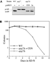
Cloning and characterization of APG7. (A) WT (wild-type, SEY6210), apg7 (THY193), apg7Δ (VDY1), and the apg7Δ strain transformed with single copy (CEN, pAPG7(414)) or multicopy (2 μ, pAPG7(424)) plasmids encoding APG7 were grown to log phase in SMD. Protein extracts were prepared and analyzed by immunoblot using antiserum to API as described in MATERIALS AND METHODS. The positions of precursor and mature API are indicated. The APG7 gene complements the precursor API accumulation phenotype of the apg7Δ mutant. (B) WT (wild-type, SEY6210), apg7Δ (VDY1), and the apg7Δ strain transformed with the APG7 centromeric plasmid pAPG7(414) were grown in SMD and transferred to SD-N as described in MATERIALS AND METHODS. Aliquots were removed at the indicated times and spread onto YPD plates in triplicate. Numbers of viable colonies were determined after 2–3 d. The APG7 gene complements the starvation-sensitive phenotype of the apg7Δ mutant.
Figure 3
Figure 3
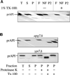
The apg7Δ mutant accumulates precursor API in a membrane-associated and protease-sensitive form that is part of a large complex. (A) The apg7Δ strain (VDY1) was grown in SMD to midlog phase and converted to spheroplasts. The spheroplasts were lysed in osmotic lysis buffer (see MATERIALS AND METHODS). An aliquot was removed for a total lysate control (T). The remainder of the lysed spheroplasts were separated into supernatant (S) and pellet (P) fractions by centrifugation at 5000 × g. The pellet fraction was resuspended in 60% sucrose in GB in the presence or absence of Triton X-100 and overlaid with 55 and 35% sucrose in GB. The step gradients were centrifuged at 100,000 × g for 60 min. Membrane-containing float (F), nonfloat (NF), and pellet (P2) fractions were collected and subjected to immunoblot analysis with antiserum to API as described in MATERIALS AND METHODS. The position of precursor API is indicated. (B) Precursor API in apg7Δ is protease accessible. Spheroplasts isolated from apg7Δ (VDY1) and ypt7Δ (WSY99) cells were lysed in osmotic lysis buffer. Supernatant (S) and pellet (P) fractions after a 5000 × g centrifugation were collected, and the pellet fractions were subjected to protease treatment in the absence or presence of 0.2% Triton X-100 as described in MATERIALS AND METHODS. The resulting samples were subjected to immunoblot analysis with antibody against API.
Figure 4
Figure 4
Apg7p is not glycosylated or proteolytically modified. (A) WT (wild-type, SEY6210), apg7Δ (VDY1) and the apg7Δ strain transformed with single-copy (CEN) or multicopy (2 μ) plasmids encoding APG7 were grown to log phase in SMD. Cells were labeled for 10 min and immunoprecipitated with antiserum to Apg7p and analyzed by SDS-PAGE as described in MATERIALS AND METHODS. Apg7p is detected as a 71-kDa protein. (B) Wild-type (SEY6210) cells were radiolabeled for 10 min and subjected to a nonradioactive chase. At the indicated time points an aliquot was removed and precipitated with TCA. Protein extracts were prepared and successively immunoprecipitated with antiserum to API and Apg7p and analyzed as above. The positions of precursor and mature API and of Apg7p are indicated. The absence of a molecular mass shift indicates that Apg7p is not proteolytically modified. (C) Wild-type (SEY6210) cells were converted to spheroplasts and treated with tunicamycin (final concentration 20 μg/ml) to inhibit glycosylation 15 min before the addition of radioactive label as indicated. Labeling was allowed to continue for 20 min. Samples were TCA precipitated and divided in half. One-half was immunoprecipitated immediately with antiserum to CPY or Apg7p for a total (T) control. The remaining half was precipitated with Con A-Sepharose and separated into supernatant (S, not bound to Con A) and pellet (P, bound to Con A) fractions as referenced in MATERIALS AND METHODS. The separate fractions were then immunoprecipitated with antiserum to CPY or Apg7p. The positions of precursor and mature forms of glycosylated CPY (p1, p2, m) and unglycosylated precursor CPY (p*) and of Apg7p are shown. Apg7p does not bind Con A, suggesting that it is not glycosylated.
Figure 5
Figure 5
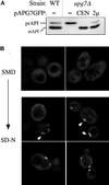
Apg7pGFP membrane association in vivo. (A) WT (wild-type, SEY6210), apg7Δ (VDY1), and the apg7Δ strain transformed with single-copy (CEN) or multicopy (2μ) plasmids encoding Apg7p-GFP were grown to log phase in SMD. Protein extracts were prepared and analyzed by Western blot using antiserum to API as described in MATERIALS AND METHODS. The positions of precursor and mature API are indicated. The Apg7pGFP hybrid protein complements the precursor API accumulation phenotype of the apg7Δ strain, indicating that the protein retains Apg7p function. (B) The apg7Δ strain transformed with the multicopy pAPG7GFP plasmid was grown in SMD to log phase and shifted to SD-N for 15 h. Cells from the SMD and SD-N cultures were examined by fluorescence microscopy as described in MATERIALS AND METHODS. Apg7pGFP displays primarily a diffuse cytosolic staining, but punctate structures are detected in SD-N medium. Some of the punctate structures appear rod-like in shape as indicated by arrows.
Figure 6
Figure 6
Apg7p subcellular fractionation pattern in SMD and SD-N. Cells from the wild-type strain (SEY6210) transformed with the multicopy plasmid encoding APG7 were grown in SMD to midlog phase and shifted to SD-N medium for 15 h before converting them to spheroplasts. The spheroplasts were then lysed osmotically in a physiological salts buffer. After a preclearing centrifugation step at 500 × g for 5 min to remove unlysed spheroplasts, the total lysate (T) was separated into 13,000 × g supernatant (S13) and pellet (P13) fractions. The S13 fraction was further separated into 100,000 × g supernatant (S100) and pellet (P100) fractions. The T, S13, P13, S100, and P100 subcellular fractions were subjected to immunoblot analysis using antiserum to Apg7p and phosphoglycerate kinase (a cytosolic marker protein). A background band (*) appears below the signal for Apg7p.
Figure 7
Figure 7
Membrane association of Apg7pGFP is dependent on Apg12p. The apg5Δ (MGY101) and apg12Δ (YNM101) strains transformed with the multicopy pAPG7GFP(426) plasmid were analyzed by fluorescence microscopy following transfer to SD-N as described in the legend to Figure 5. The punctate staining pattern of Apg7pGFP, marked by arrows, is dependent on the Apg12 protein.
Figure 8
Figure 8
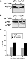
The Pichia pastoris GSA7 gene partially complements the apg7 defect. (A) The apg7Δ strain (VDY1) was transformed with a plasmid encoding the APG7 gene cloned behind the regulable CUP1 promoter (pCu416APG7). Cells were grown in YNB minus copper containing either 100 μM BCS copper chelator or 100 μM copper sulfate. After 7 h, cells were TCA precipitated, and the protein extract was analyzed by immunoblot using antiserum to Apg7p. Induction by copper results in a significant increase in Apg7p synthesis. A background band has been indicated by an asterisk (*). (B) The apg7Δ strain was transformed with a plasmid encoding the GSA7 gene cloned behind the regulable CUP1 promoter (pCu416GSA7). Cells were grown as above and analyzed by immunoblot with antiserum to API. The GSA7 gene partially complements the apg7 defect. (C) The apg7Δ strain transformed with the pCu416GSA7 or the pCu416APG7 plasmid was grown in YNB minus copper containing 100 μM coppper sulfate and transferred to SD-N as described in MATERIALS AND METHODS. Aliquots were removed at the indicated times and spread onto YPD plates in triplicate. Numbers of viable colonies were determined after 2–3 d. The pCu416GSA7 plasmid partially complements the starvation-sensitive phenotype of the apg7Δ mutant.

Similar articles

Cited by

References

    1. Baba M, Osumi M, Ohsumi Y. Analysis of the membrane structures involved in autophagy in yeast by freeze-replica method. Cell Struct Funct. 1995;20:465–471. - PubMed
    1. Baba M, Osumi M, Scott SV, Klionsky DJ, Ohsumi Y. Two distinct pathways for targeting proteins from the cytoplasm to the vacuole/lysosome. J Cell Biol. 1997;139:1687–1695. - PMC - PubMed
    1. Baba M, Takeshige K, Baba N, Ohsumi Y. Ultrastructural analysis of the autophagic process in yeast: detection of autophagosomes and their characterization. J Cell Biol. 1994;124:903–913. - PMC - PubMed
    1. Babst M, Sato TK, Banta LM, Emr SD. Endosomal transport function in yeast requires a novel AAA-type ATPase, Vps4p. EMBO J. 1997;16:1820–1831. - PMC - PubMed
    1. Baum P, Thorner J, Honig L. Identification of tubulin from the yeast Saccharomyces cerevisiae. Proc Natl Acad Sci USA. 1978;75:4962–4966. - PMC - PubMed

Publication types

MeSH terms