. 1999 May;10(5):1477-94.
doi: 10.1091/mbc.10.5.1477.

Association of myosin I alpha with endosomes and lysosomes in mammalian cells

Affiliations
Free PMC article

Association of myosin I alpha with endosomes and lysosomes in mammalian cells

G Raposo et al. Mol Biol Cell. 1999 May.
Free PMC article

Abstract

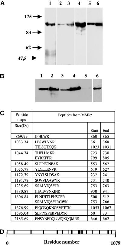
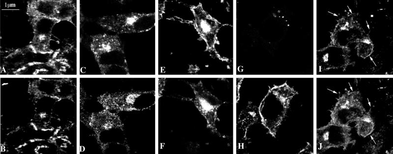
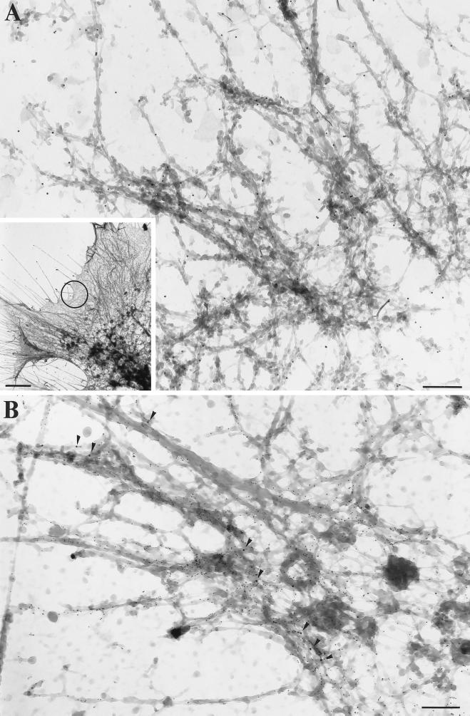
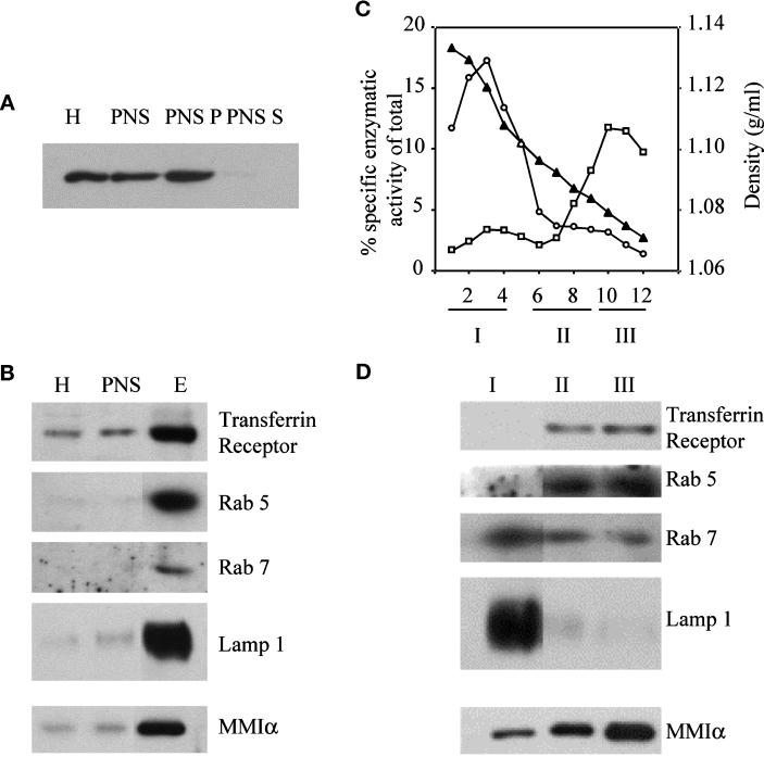
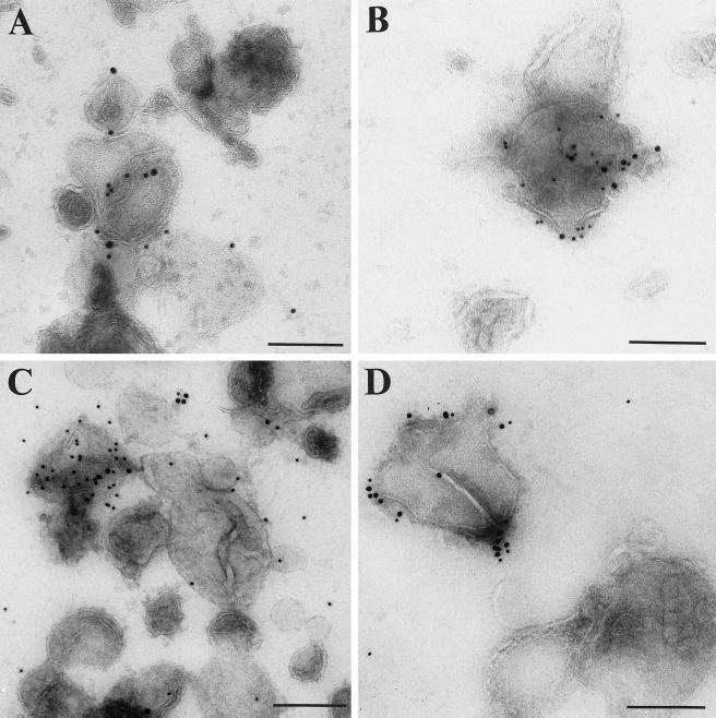
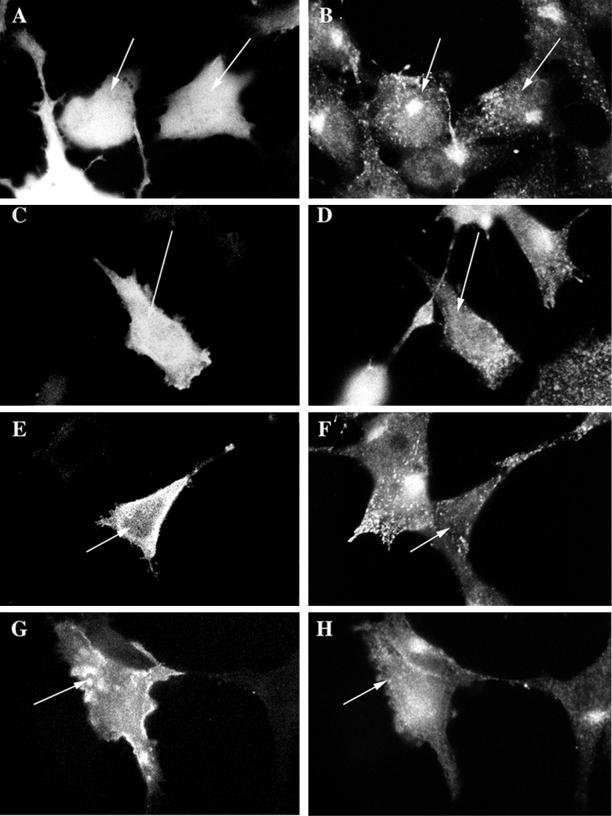
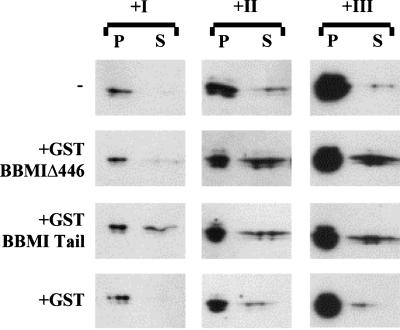
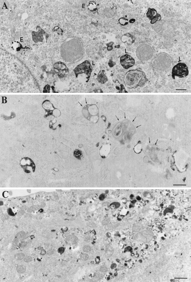
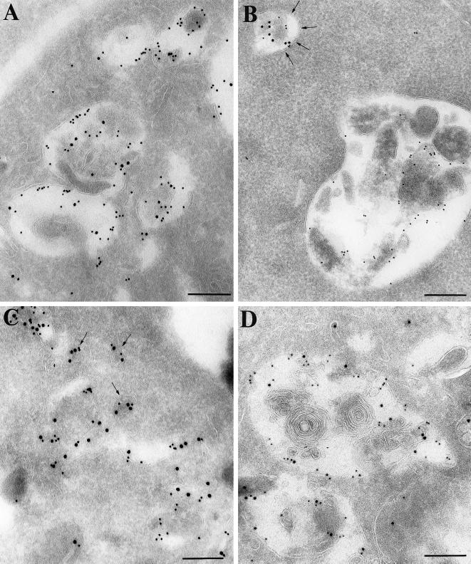
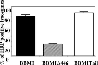
Myosin Is, which constitute a ubiquitous monomeric subclass of myosins with actin-based motor properties, are associated with plasma membrane and intracellular vesicles. Myosin Is have been proposed as key players for membrane trafficking in endocytosis or exocytosis. In the present paper we provide biochemical and immunoelectron microscopic evidence indicating that a pool of myosin I alpha (MMIalpha) is associated with endosomes and lysosomes. We show that the overproduction of MMIalpha or the production of nonfunctional truncated MMIalpha affects the distribution of the endocytic compartments. We also show that truncated brush border myosin I proteins, myosin Is that share 78% homology with MMIalpha, promote the dissociation of MMIalpha from vesicular membranes derived from endocytic compartments. The analysis at the ultrastructural level of cells producing these brush border myosin I truncated proteins shows that the delivery of the fluid phase markers from endosomes to lysosomes is impaired. MMIalpha might therefore be involved in membrane trafficking occurring between endosomes and lysosomes.


Figures

Figure 1
Figure 1
Antibodies directed against BBMI and Myr 1 recognize MMIα in the BWTG3 cells. (A) Twenty micrograms of proteins from mouse liver homogenate (lanes 1 and 4), total BWTG3 cell homogenate (lanes 2 and 5), and postnuclear supernatant (lanes 3 and 6) were separated by 7% SDS-PAGE, and transferred to nitrocellulose membranes. The membranes were probed with a monoclonal antibody directed against BBMI head domain (CX-1; lanes 1–3), or with anti-Myr 1 antibodies (Tu 30; lanes 4–6). (B) Western blot analysis of BWTG3 cell lysate immunoprecipitated with a monoclonal antibody directed against BBMI head domain (CX-1) or the antibodies directed against Myr 1 (Tu 30). Total cell lysate (lane 1), total cell lysate immunoprecipitate with anti-BBMI antibody (lane 2), supernatant of the cell lysate after the immunoprecipitation with anti-BBMI antibody (lane 3), supernatant immunoprecipitate with anti-Myr 1antibodies (Tu 30; lanes 4 and 6), and supernatant after immunoprecipitations with both antibodies (lane 5) were separated by 7% SDS-PAGE and transferred to nitrocellulose membranes. The membranes were probed with anti-Myr 1 antibodies (Tu 30; lanes 1–5) and with the anti-BBMI antibody (CX-1; lane 6). (C) The peptide map derived from the trypsin proteolysis of the 130-kDa protein immunoprecipitated with anti-Myr 1 was compared with the OWL data base using a Baysien algorithm (Profound). Thirteen peptides matched 16 peptides of MMIα. (D) Distribution of matched peptides along the sequence of MMIα. Note that the majority of the peptides were located on the tail sequence that diverges more from one subclass of myosin I to another.
Figure 2
Figure 2
Subcellular distribution of MMIα in BWTG3 cells by confocal immunofluorescence analysis. Paraformaldehyde-fixed and saponin-permeabilized cells were double labeled with phalloidin (A) and anti-Myr 1 antibodies (Tu 30; B). Cells were incubated with biotinylated transferrin 20 min before fixation, permeabilized with saponin, and double labeled with Texas Red-streptavidin (C) and anti-Myr 1 antibodies (Tu 22; D). Note that two antibodies directed against two different domains of Myr 1 decorated similarly punctate structures in the perinuclear region. After preservation of the endocytic compartments according to the procedure of Stoorvogel et al. (1996) (see MATERIALS AND METHODS), cells were paraformaldehyde fixed, detergent permeabilized, and double labeled with anti-Myr 1 antibodies (Tu 30; F, H, and J) and antibody directed against the transferrin receptor (H68.4; E), Golgi complex (G), and β actin (I). Horizontal optical sections throughout the focal plane of the nucleus were obtained by confocal laser scanning microscopy.
Figure 3
Figure 3
Visualization of MMIα and β-actin by whole-mount EM. (A) Inset, Low magnification (bar, 2 μm) of a filamentous network in the leading edge of BWTG3 cells. DAB-positive tubular and vesicular structures were observed throughout this region, but they were highly concentrated in the distal part of the leading edges. (A) Higher magnification of the circled area in inset. The anti-Myr 1 antibodies (Tu 30; PAG 10) labeled filamentous structures (A). Such a filamentous network to which MMIα (PAG 10; arrowheads) localizes was also immunogold labeled with anti-β-actin antibodies (PAG 5; panel B). (A-B) Bars, 200 nm.
Figure 4
Figure 4
Visualization of MMIα, the transferrin receptor and lgp 120 by whole-mount EM. (A) DAB-positive tubulo-vesicular structures were intensely labeled with the anti-transferrin receptor antibody (H68.4) directed against its cytoplasmic tail (PAG 10; arrows). (B) Similar DAB-positive tubulo-vesicular structures (endosomes) were labeled with the anti-Myr 1 antibodies (PAG 10, Tu 30; B, arrows). (C) Double immunogold labeling with the anti-Myr 1 antibodies (PAG 10) and the anti-transferrin receptor antibody (PAG 5) showed the codistribution of MMIα and transferrin receptor in the same tubulo-vesicular structures (arrows). (C) Inset, Lgp 120 (PAG 5) was detected in DAB-positive lysosomal compartments (arrows), which were also labeled with the anti-Myr 1 antibodies (PAG 10) (arrowheads). Bars, 200 nm.
Figure 5
Figure 5
Detection of MMIα in subcellular fractions of BWTG3 cells isolated by a sucrose step gradient or Percoll gradient. (A and B) Five micrograms of proteins from BWTG3 homogenate (H), postnuclear supernatant (PNS), and fractions collected on a three-step sucrose gradient at the interface of sucrose 1 and 0.25 M (E) were separated by 7% SDS-PAGE and transferred on nitrocellulose membranes. (A) Five micrograms of postnuclear supernatant were centrifuged 30 min at 300,000 × g. Proteins from the pellet (PNS P) and from the supernatant (PNS S) were analyzed under the same conditions. The membranes were probed with anti-Myr 1 antibodies (Tu 30; A), anti-transferrin receptor (H68.4), anti-rab 5, anti-rab 7, anti-LAMP-1, and anti-Myr 1 (Tu 30) antibodies (B). (C) Fractions collected after separation by Percoll density gradient of the postnuclear supernatant from BWTG3 cells were assayed for β-hexosaminidase (○, lysosomes), and alkaline phosphodiesterase (□, plasma membrane) activities. The linearity of percoll density after centrifugation was measured by refractometry (▴). Further analyses were always restricted to the linear part of the gradient. The graph shows enzymatic activities that correspond to the average of the activity of four fractions with the same position on four Percoll gradients performed at the same time. (D) The fractions of the four gradients were combined into three pools as indicated below the graph. Five micrograms of proteins of each pool were loaded on 7 and 10% SDS-polyacrylamide gels and analyzed by immunoblotting with the anti-transferrin receptor (H68.4), anti-rab 5, anti-rab 7, anti-LAMP-1, and anti-Myr 1 (Tu 30) antibodies. Fraction 5 was voluntarily omitted in the pools to clearly delimit pools I and II.
Figure 6
Figure 6
Immunogold labeling of endocytic compartments isolated by sucrose gradient with anti-MMIα antibodies and antibodies directed against specific markers of endocytic compartments (transferrin receptor and lgp 120). Fractions collected after separating the postnuclear supernatant from BWTG3 cells by a sucrose density gradient and enriched in endocytic markers (see Figure 5B, lane E) were loaded onto EM grids. These membrane vesicles were double immunogold labeled (A) with anti-Myr 1 antibodies (Tu 30, PAG 15) and anti-transferrin receptor antibody (PAG 10; B), with anti-Myr 1 antibodies (PAG 15) and anti-cytoplasmic tail lgp 120 antibodies (PAG 10), and (C and D) with anti-BBMI antibody (CX-1; PAG 15) and anti-β-actin antibodies (PAG 10). (D) Cells were treated with cytochalasin D before fractionation. Bars, 200 nm
Figure 7
Figure 7
The production of GFP-Myr 1, GFP-Myr 1Δn295, and GFP-Myr 1 tail affects the distribution of transferrin receptor. Twenty-four hours after transfection with cDNA encoding GFP (A and B), GFP-Myr 1 (C and D), GFP-Myr 1Δn295 (E and F), and GFP-Myr 1-Tail (G and H), cells were fixed and permeabilized with saponin. Preparations were analyzed by epifluorescent microscopy, for both the expression of the GFP recombinant proteins (A, C, E, and G) and the distribution of endosomes bearing transferrin receptor (B, D, F, and H). Cells producing GFP proteins are indicated by arrows.
Figure 8
Figure 8
In vitro competition of GST-BBMIΔ446 and GST-BBMI-Tail proteins with the MMIα associated with pools I–III. Membranes contained in the three pools were incubated in PBS at 0.5 mg/ml for 2 h at 4°C alone or with 2 μM GST-BBMIΔ446, GST-BBMI-Tail, or GST. After incubation with the fusion proteins, samples were centrifuged at 300,000 × g for 30 min. Proteins recovered from pellets and supernatants were separated by a 7% SDS-PAGE, transferred on nitrocellulose membrane, and probed with anti-MMIα antibodies (Tu 30). Samples loaded correspond to the treatment of 2.5 and 37.5 μg of total membrane proteins for the pellets and the supernatants, respectively.
Figure 9
Figure 9
Internalization of HRP by BWTG3 mock cells and cells producing BBMIΔ446 and BBMI-Tail. Cells were allowed to internalize HRP for 40 min at 37°C, fixed, and processed for DAB cytochemistry and conventional EM. In mock-treated cells (A) the electron-dense DAB reaction product was detected in intracellular compartments, which were morphologically related to endosomes (E) and lysosomes (L). In cells producing BBMIΔ446 (B), lysosomal-like compartments were devoid of electron-dense reaction product (arrows). In cells overexpressing BBMI-Tail (C), the reaction product accumulated in small vesicles distributed beneath the plasma membrane. Bars, 100 nm.
Figure 10
Figure 10
Immunogold localization of HRP (PAG 15) and cathepsin D (PAG 10) on ultrathin cryosections of BWTG3 cells producing BBMI (A), BBMIΔ446 (B), and BBMI-Tail (C and D). Before fixation cells were allowed to internalize HRP for 40 min at 37°C. In cells producing wild-type BBMI (A), the majority of cathepsin D-positive compartments contained large amounts of HRP. On the opposite, in cells producing BBMIΔ446 (B), HRP mostly accumulated in smaller compartments that were faintly labeled with the anti-cathepsin D antibodies (arrows), and the majority of lysosomes did not contain HRP. In cells overexpressing BBMI-Tail, HRP was detected in lysosomes (D) and in small cathepsin D-positive vesicular structures that were distributed at the cell periphery beneath the plasma membrane (C, arrows). Bars, 200 nm.
Figure 11
Figure 11
Semi-quantitative analysis of immunogold labeling for HRP and cathepsin D on ultrathin cryosections of cells producing BBMI, BBMIΔ446, and BBMI-Tail. Cathepsin D-positive compartments that contained more than 10 gold particles (lysosomes) were screened for the presence of HRP. Errors bars represent the SD calculated for two independent experiments.

References

    1. Bailly E, Favel J, Mahouy G, Jaureguyberry G. Plasmodium falsiparum: isolation and characterization of a 55 kDa protease with cathepsin like activity from Plasmodium falsiparum. Exp Parasitol. 1991;72:278–284. - PubMed
    1. Baker JP, Titus MA. Myosins: matching functions with motors. Curr Opin Cell Biol. 1998;10:80–86. - PubMed
    1. Bakker AC, Webster P, Jacob WA, Andrews NW. Homotypic fusion between aggregated lysosomes triggered by elevated (Ca2+) in fibroblasts. J Cell Sci. 1997;110:2227–2238. - PubMed
    1. Bearer E, DeGiorgis J, Jaffe H, Medeiros N, Reese T. An axoplasmic myosin with a calmodulin-like light chain. Proc Natl Acad Sci USA. 1996;12:6064–6068. - PMC - PubMed
    1. Bement WM, Hasson T, Wirth JA, Cheney RE, Mooseker MS. Identification and overlapping expression of multiple unconventional myosin genes in vertebrate cells types. Proc Natl Acad Sci USA. 1994;91:6549–6553. - PMC - PubMed

Publication types

MeSH terms

LinkOut - more resources