. 1999 May;10(5):1511-20.
doi: 10.1091/mbc.10.5.1511.

Radixin is involved in lamellipodial stability during nerve growth cone motility

Affiliations
Free PMC article

Radixin is involved in lamellipodial stability during nerve growth cone motility

L Castelo et al. Mol Biol Cell. 1999 May.
Free PMC article

Abstract

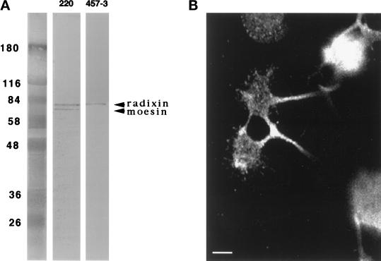
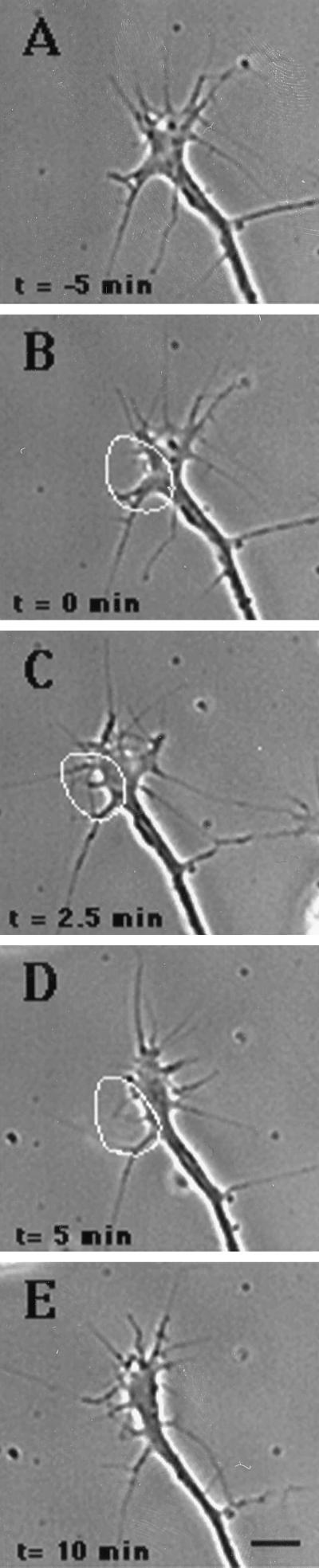
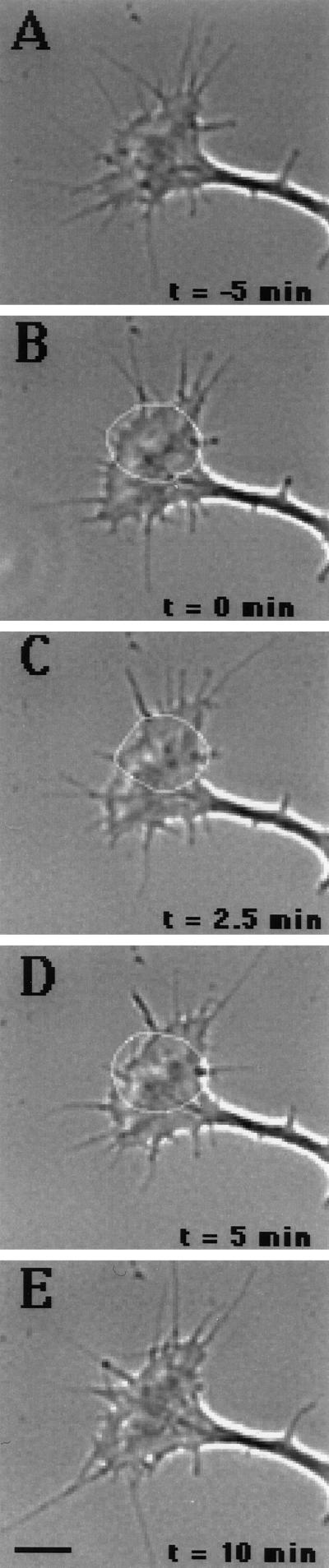
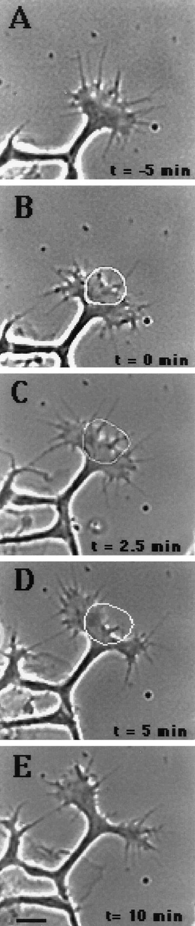
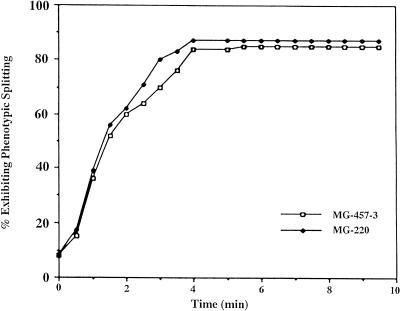
Immunocytochemistry and in vitro studies have suggested that the ERM (ezrin-radixin-moesin) protein, radixin, may have a role in nerve growth cone motility. We tested the in situ role of radixin in chick dorsal root ganglion growth cones by observing the effects of its localized and acute inactivation. Microscale chromophore-assisted laser inactivation (micro-CALI) of radixin in growth cones causes a 30% reduction of lamellipodial area within the irradiated region whereas all control treatments did not affect lamellipodia. Micro-CALI of radixin targeted to the middle of the leading edge often split growth cones to form two smaller growth cones during continued forward movement (>80%). These findings suggest a critical role for radixin in growth cone lamellipodia that is similar to ezrin function in pseudopodia of transformed fibroblasts. They are consistent with radixin linking actin filaments to each other or to the membrane during motility.


Figures

Figure 1
Figure 1
Specificity of anti-radixin antibodies. (A) DRG neuronal lysates analyzed by immunoblot with rabbit polyclonal antibodies 220 (lane a) and 457–3 (lane b). Antibody 457–3 (which is specific for radixin) recognizes a single band corresponding to radixin. Antibody 220 (which binds to all three ERM proteins) recognizes two bands with molecular weights corresponding to radixin and moesin. DRG neuronal lysates do not appear to express ezrin. (B) DRG-dissociated neurons were grown on laminin-poly-l-lysine–coated coverslips and were fixed and probed with 457–3 anti-radixin followed by FITC-labeled anti-rabbit IgG. Radixin is present throughout the growth cone and exhibits a punctate pattern. Staining was undetectable for controls that lack 457–3.
Figure 2
Figure 2
Micro-CALI of radixin causes lamellipodial retraction. A chick DRG neuron was loaded with MG-labeled 457–3 and plated on a laminin-coated coverslip to allow a growth cone to emerge and time-lapse imaging was begun at t = −5 min (A). The growth conewas subjected to laser irradiation on one side of the growth cone (white circle) beginning at t = 0 min (B) and ending at t = 5 min (C), and the growth cone was observed for another 10 min (D and E). The lamellipodia in the irradiated half retracts (B–D) and begins to recover by t = 10 min (E). Scale bar, 10 μm.
Figure 3
Figure 3
Micro-CALI using MG-nonimmune IgG has no effect. A chick DRG neuron was loaded with MG-labeled nonimmune IgG and plated on laminin-coated coverslip to allow a growth cone to emerge. Time-lapse imaging was begun at t = −5 min (A). The growth cone was subjected to laser irradiation within a small region of the growth cone (white circle) beginning at t = 0 min (B) andending at t = 5 min (C), and the growth cone was observed for another 10 min (D and E). The lamellipodia within the irradiated region do not retract (B–E). Scale bar, 10 μm.qending at t = 5 min (C), and the growth cone was observed for another 10 min (D and E). The lamellipodia within the irradiated region do not retract (B–E). Scale bar, 10 μm.
Figure 4
Figure 4
Micro-CALI of radixin in the central region causes growth cone splitting. A chick DRG neuron was loaded with MG-labeled 457–3 and plated on a laminin-coated coverslip to allow agrowth cone to emerge. Time-lapse imaging was begun at t = −5 min (A). The growth cone was subjected to laser irradiation in its central region (white circle) beginning at t = 0 min (B) and ending at t = 5 min (C), and the growth cone was observed for another 10 min (D and E). The lamellipodia in the irradiated region retract (B and C), and the growth cone splits into two small growth cones (D and E). Similar results were seen using MG-labeled 220 in place of MG-labeled 457–3 (our unpublished results). No effect was seen with MG-labeled nonimmune IgG (see Table 2). Scale bar, 10 μm.
Figure 5
Figure 5
The effects of micro-CALI of radixin and of all ERMs are indistinguishable. The time course for growth cone splitting was plotted for all samples subjected to micro-CALI of radixin alone and micro-CALI of all ERMs. Growth cone splitting is initiated early during laser irradiation and occurs rapidly for both treatments.

References

    1. Algrain M, Turunen O, Vaheri A, Louvard D, Arpin M. Ezrin contains cytoskeletal and membrane binding domains accounting for its proposed role as a membrane-cytoskeletal linker. J Cell Biol. 1993;120:129–139. - PMC - PubMed
    1. Arpin M, Algrain M, Louvard D. Membrane-actin microfilament connection: an increasing diversity of players related to band 4.1. Curr Opin Cell Biol. 1994;6:136–141. - PubMed
    1. Beerman AE, Jay DG. Chromophore-assisted laser inactivation of cellular proteins. Methods Cell Biol. 1994;44:716–732. - PubMed
    1. Bentley D, Toroian-Raymond A. Disoriented pathfinding by pioneer neuron growth cones deprived of filopodia by cytochalasin treatment. Nature. 1986;323:712–715. - PubMed
    1. Birgbauer E, Dinsmore JH, Winckler B, Lander AD, Solomon F. Association of ezrin isoforms with the neuronal cytoskeleton. J Neurosci Res. 1991;30:232–241. - PubMed

Publication types