. 1999 May;10(5):1621-36.
doi: 10.1091/mbc.10.5.1621.

Endocytosis deficiency of epidermal growth factor (EGF) receptor-ErbB2 heterodimers in response to EGF stimulation

Affiliations
Free PMC article

Endocytosis deficiency of epidermal growth factor (EGF) receptor-ErbB2 heterodimers in response to EGF stimulation

Z Wang et al. Mol Biol Cell. 1999 May.
Free PMC article

Abstract

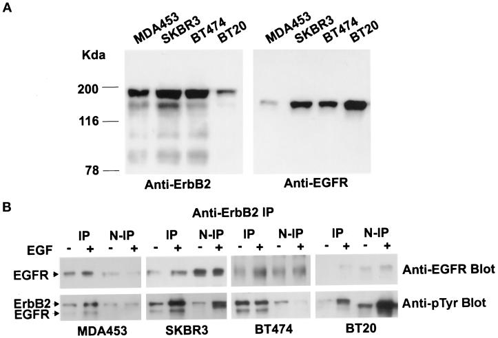
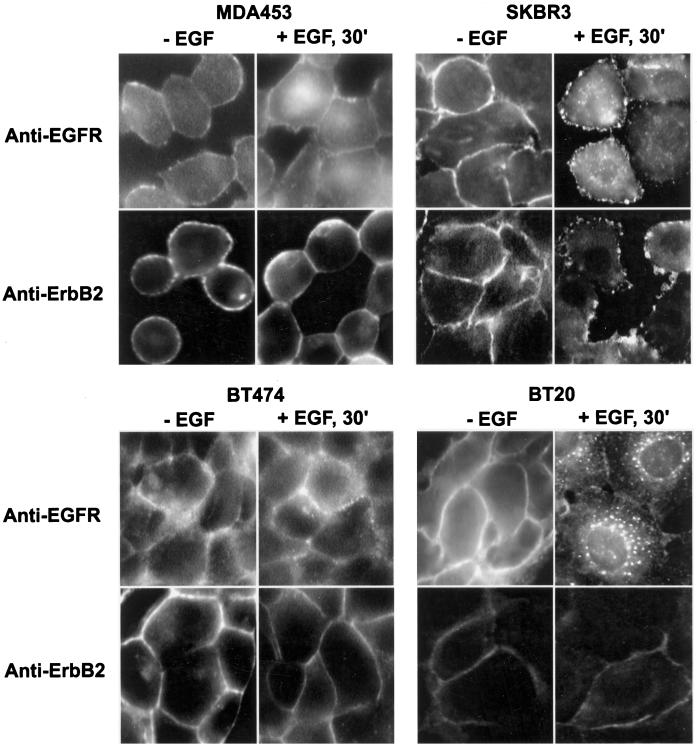
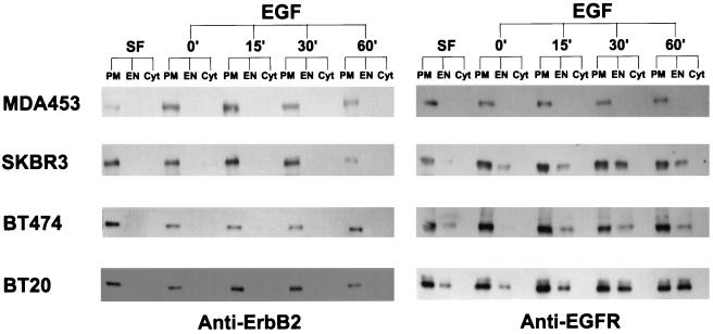
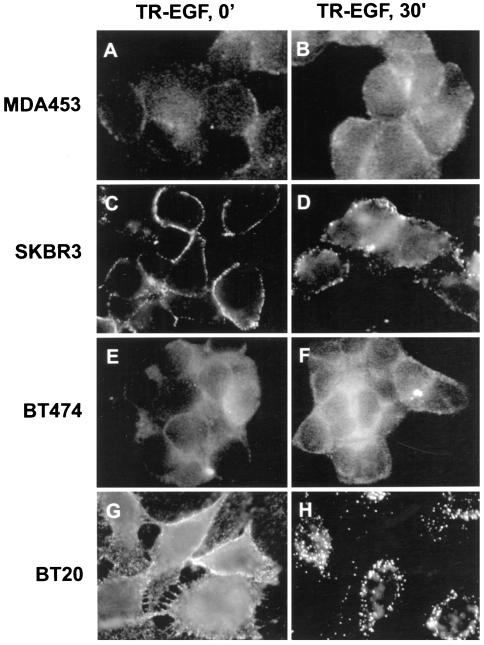
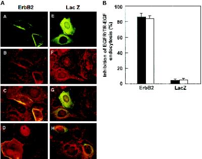
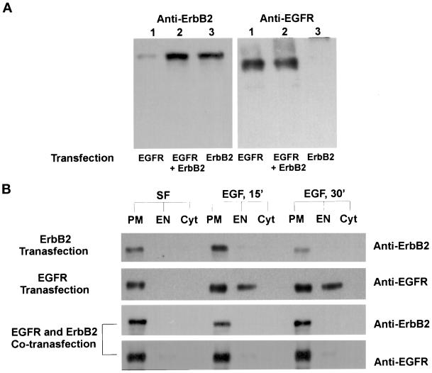
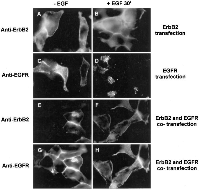
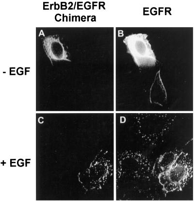
Epidermal growth factor (EGF) stimulates the homodimerization of EGF receptor (EGFR) and the heterodimerization of EGFR and ErbB2. The EGFR homodimers are quickly endocytosed after EGF stimulation as a means of down-regulation. However, the results from experiments on the ability of ErbB2 to undergo ligand-induced endocytosis are very controversial. It is unclear how the EGFR-ErbB2 heterodimers might behave. In this research, we showed by subcellular fractionation, immunoprecipitation, Western blotting, indirect immunofluorescence, and microinjection that, in the four breast cancer cell lines MDA453, SKBR3, BT474, and BT20, the EGFR-ErbB2 heterodimerization levels were positively correlated with the ratio of ErbB2/EGFR expression levels. ErbB2 was not endocytosed in response to EGF stimulation. Moreover, in MDA453, SKBR3, and BT474 cells, which have very high levels of EGFR-ErbB2 heterodimerization, EGF-induced EGFR endocytosis was greatly inhibited compared with that in BT20 cells, which have a very low level of EGFR-ErbB2 heterodimerization. Microinjection of an ErbB2 expression plasmid into BT20 cells significantly inhibited EGF-stimulated EGFR endocytosis. Coexpression of ErbB2 with EGFR in 293T cells also significantly inhibited EGF-stimulated EGFR endocytosis. EGF did not stimulate the endocytosis of ectopically expressed ErbB2 in BT20 and 293T cells. These results indicate that ErbB2 and the EGFR-ErbB2 heterodimers are impaired in EGF-induced endocytosis. Moreover, when expressed in BT20 cells by microinjection, a chimeric receptor composed of the ErbB2 extracellular domain and the EGFR intracellular domain underwent normal endocytosis in response to EGF, and this chimera did not block EGF-induced EGFR endocytosis. Thus, the endocytosis deficiency of ErbB2 is due to the sequence of its intracellular domain.


Figures

Figure 1
Figure 1
EGFR and ErbB2 concentrations, EGF-stimulated heterodimerization, and phosphorylation of EGFR and ErbB2 in various breast cancer cell lines. (A) MDA453, SKBR3, BT474, and BT20 cells were lysed with radioimmunoprecipitation assay buffer, and 20 μg of protein from each lysate were separated by 10% SDS-PAGE, transferred onto a nitrocellulose filter, and incubated with rabbit polyclonal anti-ErbB2 or anti-EGFR antibody. (B) MDA453, SKBR3, BT474, and BT20 cells were serum starved or stimulated with EGF (100 ng/ml) at 4°C for 60 min and then lysed with immunoprecipitation buffer. The total lysates were immunoprecipitated with monoclonal anti-ErbB2 antibody. Then, formula image of the total protein from both the anti-ErbB2 immunoprecipitates and the nonprecipitated supernatants was separated by 10% SDS-PAGE, transferred onto a nitrocellulose filter, and incubated with a rabbit polyclonal anti-EGFR antibody or a mouse monoclonal anti-pTyr antibody. Bands were visualized on x-ray film by using an HRP-conjugated secondary antibody and chemiluminescence reagents.
Figure 2
Figure 2
Determination by immunofluorescence analysis of ErbB2 and EGFR endocytosis after EGF stimulation. MDA453, SKBR3, BT474, and BT20 cells on glass coverslips were serum starved or stimulated with EGF (100 ng/ml) at 4°C for 60 min followed by further incubation at 37°C for 30 min. The cells were fixed with methanol and stained with mouse monoclonal anti-ErbB2 or anti-EGFR antibody followed with FITC-conjugated secondary antibody, as described in MATERIALS AND METHODS. Magnification, 200×.
Figure 3
Figure 3
Analysis by subcellular fractionation and immunoblotting of EGFR and ErbB2 endocytosis after EGF stimulation. MDA453, SKBR3, BT474, and BT20 cells were cultured in serum-free medium or incubated with EGF (100 ng/ml) at 4°C for 60 min, followed by further incubation with EGF at 37°C for 15, 30, and 60 min as indicated. The cells were fractionated into PM, EN, and Cyt fractions as described in MATERIALS AND METHODS. Proteins (10 μg) from each fraction were separated by SDS-PAGE, transferred onto a nitrocellulose filter, and incubated with rabbit polyclonal anti-ErbB2 or anti-EGFR antibody. Bands were visualized on x-ray film by using an HRP-conjugated secondary antibody and chemiluminescence reagents.
Figure 4
Figure 4
Determination by immunofluorescence analysis of TR-EGF internalization. MDA453, SKBR3, BT474, and BT20 cells grown on glass coverslips were incubated with TR-EGF (100 ng/ml) at 4°C for 60 min (referred as 0 min; A, C, E, and G). Some coverslips were further incubated at 37°C for 30 min (B, D, F, and H). The cells were then fixed with methanol and examined under the microscope. Magnification, 180×.
Figure 5
Figure 5
Inhibition of EGFR endocytosis in BT20 cells after microinjection of the ErbB2 expression plasmid. (A) BT20 cells were microinjected with the ErbB2 expression plasmid pcDNA3.1(−)/ErbB2 (A–D) or the control plasmid pcDNA3.1(−)/Myc-His/LacZ (E–H) and incubated at 37°C for 24 h. After treatment with EGF (100 ng/ml; (A–C and E–G) or TR-EGF (100 ng/ml; D and H), the cells were fixed with methanol and stained by immunofluorescence. ErbB2 and LacZ expression-positive cells were identified with a mouse anti-ErbB2 (A, C, and D) or mouse anti-myc (B, G, and H) antibody followed by FITC-conjugated anti-mouse IgG. EGFR endocytosis was assayed with sheep polyclonal anti-EGFR antibody followed by a TRITC-conjugated anti-sheep antibody (B, C, F, and G). The endocytosis of TR-EGF was directly examined under fluorescent microscope (D and H). Magnification, 200×. (B) Quantification of inhibition of EGFR and TR-EGF endocytosis after microinjection of ErbB2 expression plasmid. Data are means ± SE of three independent experiments.
Figure 6
Figure 6
Analysis by subcellular fractionation and immunoblotting of EGFR and ErbB2 endocytosis after EGF stimulation in the transiently transfected 293T cells. (A) Western blot analysis of EGFR and ErbB2 expression in 293T cells transiently transfected with pcDNA3.1(−)/EGFR and/or pcDNA3.1(−)/ErbB2. (B) Determination of EGF-induced EGFR and ErbB2 endocytosis in 293T cells transfected with pcDNA3.1(−)/EGFR and/or pcDNA3.1(−)/ErbB2. 293T cells were cultured in serum-free medium or stimulated with EGF at 4°C for 60 min followed by further incubation in serum-free medium for 15 or 30 min. The cells were subcellular fractionated and immunoblotted for EGFR and/or ErbB2 as described in MATERIALS AND METHODS.
Figure 7
Figure 7
Determination by immunofluorescence analysis of EGF-induced EGFR and ErbB2 endocytosis in the transiently transfected 293T cells. 293T were transiently transfected with pcDNA3.1(−)/ErbB2 (A and B), pcDNA3.1(−)/EGFR (C and D), or both (E–H). After the transfection, the cells were cultured in serum-free medium (A, C, E, and G) or incubated with EGF (100 ng/ml) at 4°C for 60 min followed by further incubation for 30 min at 37°C (B, D, F, and H). The cells were then single immunofluorescent stained for ErbB2 (A and B) and EGFR (C and D) or double immunofluorescent stained for both ErbB2 (E and F) and EGFR (G and H), as described in MATERIALS AND METHODS. Magnification, 200×.
Figure 8
Figure 8
Determination by immunofluorescence analysis of EGF-induced endocytosis of an ErbB2/EGFR chimera. BT20 cells were microinjected with the chimeric ErbB2/EGFR expression plasmid pcDNA3.1(−)/ErbB2/EGFR/Myc/His and incubated at 37°C for 24 h. The cells were either untreated (A and B) or treated with EGF (100 ng/ml) at 37°C for 30 min (C and D). The cells were then fixed with methanol and stained by immunofluorescence. The endocytosis of the chimeric ErbB2/EGFR was assayed by mouse anti-myc antibody (B, G, and H) followed by FITC-conjugated anti-mouse IgG (A and C). The EGFR endocytosis was assayed with sheep polyclonal anti-EGFR antibody followed by a TRITC-conjugated anti-sheep antibody (B and D). Magnification, 200×.

References

    1. Anders RA, Arline SL, Dore JJ, Leof EB. Distinct endocytic responses of heteromeric and homomeric transforming growth factor beta receptors. Mol Biol Cell. 1997;8:2133–2143. - PMC - PubMed
    1. Anderson D, Koch CA, Gray L, Ellis C, Moran MF, Pawson T. Binding of SH2 domains of phospholipase C gamma 1, GAP, and Src to activated growth factor receptors. Science. 1990;250:979–982. - PubMed
    1. Bargmann CI, Hung MC, Weinberg RA. Multiple independent activations of the neu oncogene by a point mutation altering the transmembrane domain of p185. Cell. 1986;45:649–657. - PubMed
    1. Baulida J, Kraus MH, Alimandi M, Di Fiore PP, Carpenter G. All ErbB receptors other than the epidermal growth factor receptor are endocytosis impaired. J Biol Chem. 1996;271:5251–5257. - PubMed
    1. Carpenter G. Receptors for epidermal growth factor and other polypeptide mitogens. Annu Rev Biochem. 1987;56:881–914. - PubMed

Publication types

MeSH terms