

The protective role of T-cell receptor Vgamma1+ T cells in primary infection with Listeria monocytogenes
- PMID: 10233675
- PMCID: PMC2326708
- DOI: 10.1046/j.1365-2567.1999.00666.x
The protective role of T-cell receptor Vgamma1+ T cells in primary infection with Listeria monocytogenes
Abstract
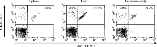
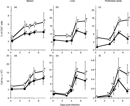
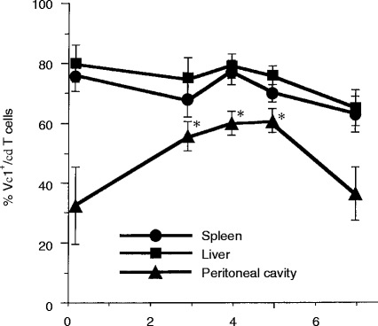
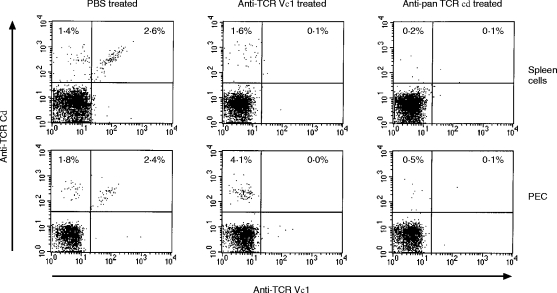
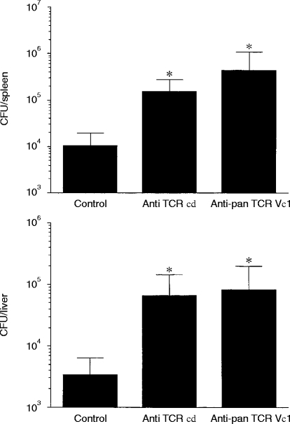
We have previously reported that the T-cell receptor (TCR) gamma delta+ T cells increase in mice infected with an intracellular bacteria Listeria monocytogenes, and the cells predominantly express Vdelta6 and Vgamma1 genes. In this study, we used a monoclonal antibody (mAb) specific to TCR Vgamma1 to estimate the frequency of Vgamma1+ T cells and we discuss their significance in protection against L. monocytogenes. The spleen, liver and peritoneal exudate cells from mice intraperitoneally infected with L. monocytogenes were analysed by flow cytometry. In all the organs investigated, Vgamma1+ cells increased predominantly among TCR gamma delta+ T cells at an early phase (day 5-7) of the infection. To elucidate the significance of the Vgamma1+ T cells in the protection against L. monocytogenes, mice were depleted of TCR Vgamma1+ gamma delta T cells or all TCR gamma delta+ T cells by intraperitoneal inoculation of anti-Vgamma1 mAb or anti-pan TCR gamma delta mAb, respectively, before infection with L. monocytogenes. The bacterial growth in the spleen and the liver examined on day 5 after the infection increased significantly by the depletion of TCR Vgamma1+ T cells. The numbers of L. monocytogenes in TCR Vgamma1+ T-cell-depleted mice were nearly the same as in mice depleted of all TCR gamma delta+ T cells. These results demonstrated that Vgamma1+ T cells are the predominant population of gamma delta T cells in protection against L. monocytogenes at the early phase of the primary infection.
Figures
Similar articles
-
Mechanism of murine Vgamma1+ gamma delta T cell-mediated innate immune response against Listeria monocytogenes infection.Eur J Immunol. 2002 Apr;32(4):928-35. doi: 10.1002/1521-4141(200204)32:4<928::AID-IMMU928>3.0.CO;2-I. Eur J Immunol. 2002. PMID: 11920558
-
Analysis of the role of natural killer cells in Listeria monocytogenes infection: relation between natural killer cells and T-cell receptor gamma delta T cells in the host defence mechanism at the early stage of infection.Immunology. 1994 May;82(1):106-12. Immunology. 1994. PMID: 8045587 Free PMC article.
-
A protective role of gamma/delta T cells in primary infection with Listeria monocytogenes in mice.J Exp Med. 1992 Jan 1;175(1):49-56. doi: 10.1084/jem.175.1.49. J Exp Med. 1992. PMID: 1530961 Free PMC article.
-
The role of gammadelta T cells in induction of bacterial antigen-specific protective CD8+ cytotoxic T cells in immune response against the intracellular bacteria Listeria monocytogenes.Immunology. 1998 Oct;95(2):226-33. doi: 10.1046/j.1365-2567.1998.00593.x. Immunology. 1998. PMID: 9824480 Free PMC article.
-
Early host-pathogen interactions in the liver and spleen during systemic murine listeriosis: an overview.Immunobiology. 1999 Dec;201(2):178-87. doi: 10.1016/S0171-2985(99)80057-6. Immunobiology. 1999. PMID: 10631566 Review.
Cited by
-
Protective role of gammadelta T cells in spontaneous ocular inflammation.Invest Ophthalmol Vis Sci. 2009 Jul;50(7):3266-74. doi: 10.1167/iovs.08-2982. Epub 2009 Jan 17. Invest Ophthalmol Vis Sci. 2009. PMID: 19151391 Free PMC article.
-
Cytokine production by Vgamma(+)-T-cell subsets is an important factor determining CD4(+)-Th-cell phenotype and susceptibility of BALB/c mice to coxsackievirus B3-induced myocarditis.J Virol. 2001 Jul;75(13):5860-9. doi: 10.1128/JVI.75.13.5860-5869.2001. J Virol. 2001. PMID: 11390587 Free PMC article.
-
Importance of murine Vdelta1gammadelta T cells expressing interferon-gamma and interleukin-17A in innate protection against Listeria monocytogenes infection.Immunology. 2008 Oct;125(2):170-7. doi: 10.1111/j.1365-2567.2008.02841.x. Epub 2008 Apr 7. Immunology. 2008. PMID: 18397272 Free PMC article.
-
From Host Defense to Metabolic Signatures: Unveiling the Role of γδ T Cells in Bacterial Infections.Biomolecules. 2024 Feb 15;14(2):225. doi: 10.3390/biom14020225. Biomolecules. 2024. PMID: 38397462 Free PMC article. Review.
-
A defective Th1 response of the spleen in the initial phase may explain why splenectomy helps prevent a Listeria infection.Clin Exp Immunol. 2005 Apr;140(1):11-21. doi: 10.1111/j.1365-2249.2005.02735.x. Clin Exp Immunol. 2005. PMID: 15762870 Free PMC article.
References
-
- Haas W, Pereira P, Tonegawa S. Gamma/delta cells. Annu Rev Immunol. 1993;11:637. - PubMed
-
- Chien YH, Jores R, Crowley MP. Recognition by γ/δ T cells. Annu Rev Immunol. 1996;14:511. - PubMed
-
- Mombaerts P, Arnoldi J, Russ F, Tonegawa S, Kaufmann SH. Different roles of αβ and γδ T cells in immunity against an intracellular bacterial pathogen. Nature. 1993;365:53. - PubMed
MeSH terms
Substances
LinkOut - more resources
Full Text Sources
Medical
