. 1999 Jan;96(1):35-47.
doi: 10.1046/j.1365-2567.1999.00670.x.

Molecular characterization of U937-dependent T-cell co-stimulation

Affiliations

Molecular characterization of U937-dependent T-cell co-stimulation

T J Stonehouse et al. Immunology. 1999 Jan.

Abstract

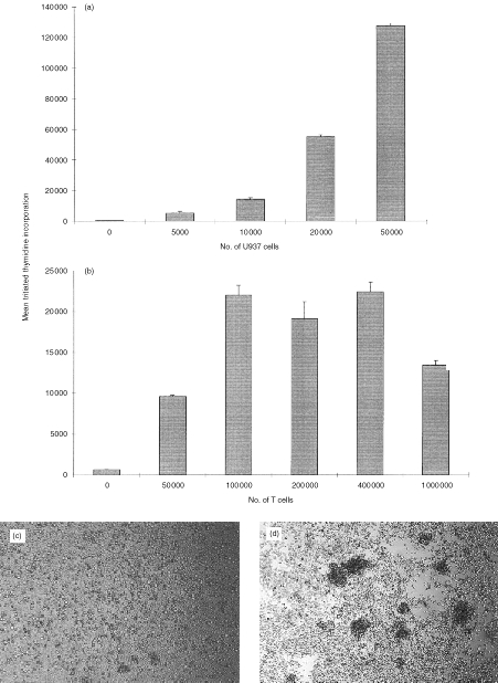
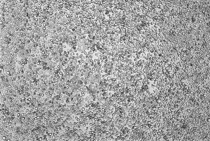
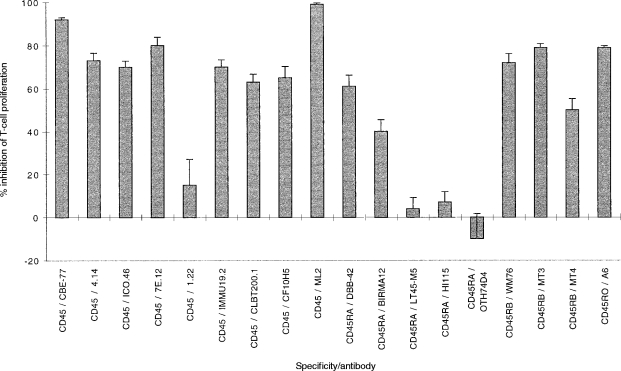
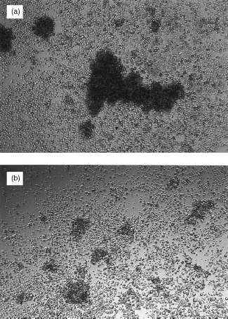
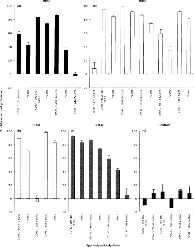
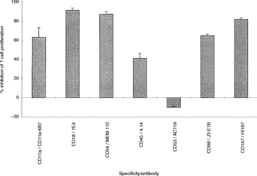
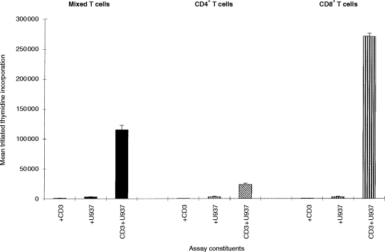
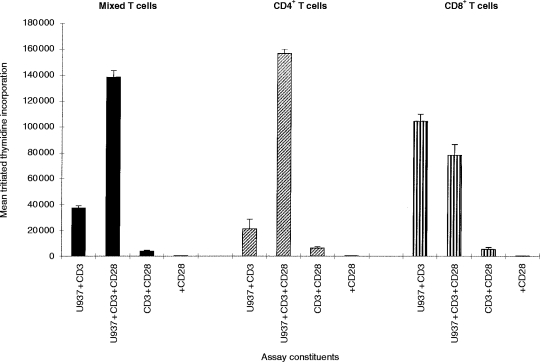
U937 cells provide a co-stimulatory signal for CD3-mediated T-cell activation which is independent of the CD28/CD80/CD86 interaction. This study set out to identify which molecules contribute to this co-stimulatory activity. Monoclonal antibodies (mAb) to the known accessory molecules CD11a, CD18, CD54 and CD45, all inhibited T-cell proliferation. Although CD11a/18 mAb inhibited U937/T-cell cluster formation as well as proliferation, CD45 enhanced the size of the clusters formed, suggesting that this was not the only mechanism of inhibition. The alternative co-stimulatory pathway provided by U937 cells preferentially stimulated a response in the CD18+ T-cell population, and this reflected the reduced sensitivity of CD8+ T cells to CD28-mediated activation. Monoclonal antibodies to three molecules, CD53, CD98 and CD147, also inhibited U937-dependent T-cell proliferation. The mAb to CD98 and CD147 were inhibitory when prepulsed on to the U937 cells, suggesting an effect mediated by these molecules on the antigen-presenting cell.


Figures

Figure 1
Figure 1
U937 cells co-stimulate T cells in a CD3-dependent proliferation assay. (a) Increasing numbers of irradiated U937 cells were co-cultured with 2×10⁵ purified tonsillar T cells, in the presence of 0·1 μg/ml CD3 mAb (UCHT1). (b) Increasing numbers of T cells were co-cultured with 104 irradiated U937 cells, in the presence of 0·1 μg/ml CD3 mAb (UCHT1). T-cell proliferation in both (a) and (b) was assessed by [3H]thymidine incorporation between 48 and 64 hr of culture. T cells and U937 together without CD3 mAb, and T cells with CD3 mAb but no U937 cells did not elicit a T-cell response. 5×10⁴ irradiated U937 cells alone gave thymidine incorporation values of 2000 c.p.m. The values shown are the mean [3H]thymidine incorporation of triplicate wells±SD from one experiment. Similar results have been found in at least two other experiments. (c) and (d) Typical U937 – T-cell clusters were seen at ×40 magnification, approximately 48 hr after the start of the assay. (c) 2×10⁵ purified tonsillar T cells, and 104 irradiated U937 cells. (d) As for (c) but with CD3 mAb (0·1 μg/ml).
Figure 2
Figure 2
The CD18 antibody 7E4 inhibits the formation of U937 – T-cell clusters. CD18 mAb 7E4 (1/400 dilution) was added to 104 irradiated U937 cells, 2×10⁵ purified tonsillar T cells, and 0·1 μg/ml of CD3 mAb (UCHT1) and cluster formation was assessed visually at ×40 magnification. Similar results were obtained with other CD11a, CD54 and other CD18 mAb.
Figure 3
Figure 3
CD45 antibodies inhibit the T-cell response. A panel of CD45 mAb were added to 104 irradiated U937 cells, 2×10⁵ purified T cells and 0·1 μg/ml CD3 mAb (UCHT1). T-cell proliferation was assessed between 48 and 64 hr of culture by measurement of [3H]thymidine incorporation. The inhibitory response is shown as the mean percentage inhibition of proliferation in triplicate wells ±% error, when test antibody is added to the assay compared to proliferation in the absence of test antibody. Similar results have been obtained in at least two other experiments.
Figure 4
Figure 4
The CD45 antibody 4.14 causes the formation of large U937–T-cell clusters. CD45 mAb 4.14 (1/400 dilution) was added to 104 irradiated U937 cells, 2×10⁵ purified T cells and 0·1 μg/ml CD3 mAb (UCHT1) (a). The cells were cultured for approximately 48 hr and then photographed. In (b) the culture conditions were the same but no CD3 mAb was added. Other inhibitory CD45 mAb (see Fig. 3) had the same effect of increasing U937 – T-cell clustering, and this increase was independent of CD45 isotype.
Figure 5
Figure 5
(a) CD53, (b) CD98 and (c) CD147 inhibit the T-cell response whereas (d) controls do not. Test mAb (final volume 1 μl mAb:200 μl) were added to cultures of 104 irradiated U937 cells, 2×10⁵ purified T cells and 0·1 μg/ml CD3 mAb (UCHT1). Proliferation was assessed between 48 and 64 hr of culture by measurement of [3H]thymidine incorporation. The response is shown as the mean percentage inhibition of T-cell proliferation in triplicate wells ±% error, when test mAb is added to the assay compared to the proliferation in the absence of test mAb. The proliferation in the absence of test mAb was 49 876 c.p.m. Similar results have been obtained in at least two other experiments.
Figure 6
Figure 6
Inhibitory activity of antibodies when bound selectively to U937 cells. Monoclonal antibodies were incubated with U937 cells for 1 hr at 37°. The mAb concentrations for these experiments were double those used for the assays outlined previously, i.e. the same protein concentration of mAb was used as before, but the dilution was 1:100 rather than 1:200. Excess mAb was removed by repeated washing, the cells were irradiated, and then 104 of U937/mAb complex were co-cultured with 2×10⁵ purified T cells and 0·1 μg/ml CD3 mAb (UCHT1). Proliferation was assessed between 48 and 64 hr of culture by measurement of [3H]thymidine incorporation. The results are expressed as the mean percentage inhibition of T-cell proliferation in triplicate wells ±% error, when test mAb was added to the U937 cells compared to the proliferation in the absence of test mAb. Similar results have been obtained in at least two other experiments.
Figure 7
Figure 7
U937 cells co-stimulate predominantly CD8+ T cells in the CD3-dependent proliferation assay. U937cells (irradiated, 104) were in the presence of co-cultured with 2×10⁵ T cells, purified CD4+ T cells, or purified CD8+ T cells (prepared as outlined in the Materials and Methods) and 0·1 μg/ml CD3 mAb (UCHT1). Proliferation was assessed between 48 and 64 hr of culture by [3H]thymidine incorporation between 48 and 64 hr of culture. The results are expressed as the mean percentage inhibition of T-cell proliferation in triplicate wells ±% error. Similar results have been obtained in at least two other experiments.
Figure 8
Figure 8
An agonistic CD28 antibody co-stimulates CD4+ T cells, but has no effect on CD8+ T cells. The CD28 mAb 15E9 (1/20) was added to 104 irradiated U937 cells, either 2×10⁵ purified T cells, or 2×10⁵ purified CD4+ or 2×10⁵ purified CD8+ T cells, and 0·1 μg/ml CD3 mAb (UCHT1). T-cell proliferation was assessed between 48 and 64 hr of culture by measurement of [3H]thymidine incorporation. The results are expressed as the mean percentage inhibition of T-cell proliferation in triplicate wells ±% error. Similar results have been found in at least two other experiments.

Similar articles

Cited by

References

    1. Schwartz RH. Costimulation of T lymphocytes: the role of CD28, CTLA-4, and B7/BB1 in interleukin-2 production and immunotherapy. Cell. 1992;71:1065. - PubMed
    1. Schwartz RH. T-lymphocyte recognition of antigen in association with gene products of the major histocompatibility complex. Annu Rev Immunol. 1985;3:237. - PubMed
    1. Barber EK, Dasgupta JD, Schlossman SF, Trevillyan JM, Rudd CE. The CD4 and CD8 antigens are coupled to a protein-tyrosine kinase (p56lck) that phosphorylates the CD3 complex. Proc Natl Acad Sci USA. 1989;86:3277. - PMC - PubMed
    1. Weiss A, Iwashima M, Irving B, et al. Molecular and genetic insights into T cell antigen receptor signal transduction. Adv Exp Med Biol. 1994;365:53. - PubMed
    1. Jenkins MK, Johnson JG. Molecules involved in T-cell costimulation. Curr Opin Immunol. 1993;5:361. - PubMed

Publication types

MeSH terms