

Review
doi: 10.1046/j.1365-2567.1999.00681.x.
Local immune responses in afferent and efferent lymph
Affiliations
- PMID: 10233690
- PMCID: PMC2326739
- DOI: 10.1046/j.1365-2567.1999.00681.x
Item in Clipboard
Review
Local immune responses in afferent and efferent lymph
Immunology.
1999 Feb.
No abstract available
Figures
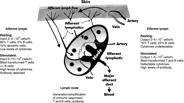
Flow rates and cellular composition of lymph around single skin-draining lymph nodes of adult sheep. Afferent lymph contains antigen-presenting dendritic cells, T cells and a small proportion of B cells. Lymph flow and cell output rates are low. Efferent lymph contains >98% lymphocytes with a higher proportion of B cells and much higher flow and cell output rates than in afferent lymph.2–5 Greater than 90% of efferent lymphocytes are derived from the blood via high endothelial venules (HEV). L-selectin on lymphocytes is associated with adhesion to peripheral lymph node HEV. For the details of the cytokines studied to date, see Fig. 2 and the text.
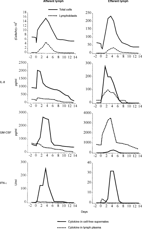
Differential cytokine production in afferent and efferent lymph during an anamnestic immune response to 1 mg orf virus antigen given intra-dermally to previously infected sheep (refs 14, 21 and unpublished). Mean total and lymphoblast output for each group of five sheep is shown for comparison (top graphs). Lymph plasma contains activity generated in the skin (afferent lymph) or prefemoral lymph node (efferent lymph) as well as by cells in the lymph. Cytokine production by 4×10⁶ lymph cells in 1 ml medium after culture for 18 hr in the absence of exogenous antigen was assayed in cell-free supernates (CFS) as a measure of the activation status of the cells following in vivo antigen challenge. Three examples of cytokines are shown. Note the difference in y-axis scales between afferent and efferent lymph. The chemokine IL-8 is produced within 12 hr of antigen challenge in both lymph compartments, most notably by afferent lymph cells. Low titres were detected in lymph plasma (there is no evidence for inhibitors in lymph plasma). GM-CSF is a haemopoietin active on macrophages, neutrophils and dendritic cells. Large quantities are produced within the lymph node and by afferent but not efferent lymph cells. This might indicate a consequence of accessory cell–lymphocyte interactions in afferent lymph and the lymph node. IFN-γ, like GM-CSF, is produced rapidly and at high titre by afferent lymph cells compared to efferent lymph cells where production is low and delayed by comparison. Production by cells correlated with the appearance of lymphoblasts. Differential and temporal appearance of the cytokines in the lymph compartments was similar in sheep reinfected with orf virus where CD4+/CD45RA− T lymphoblasts were the principal source of GM-CSF, IFN-γ (and IL-2).14,21 The results show a marked concentration of activated cytokine-producing cells in afferent lymph compared to efferent lymph during the anamnestic immune response, consistent with an activated effector/memory cell population in afferent lymph. A function for IL-8 (which precedes IL-1β detection14) in the initial phase of the anamnestic immune/inflammatory response is also implied.
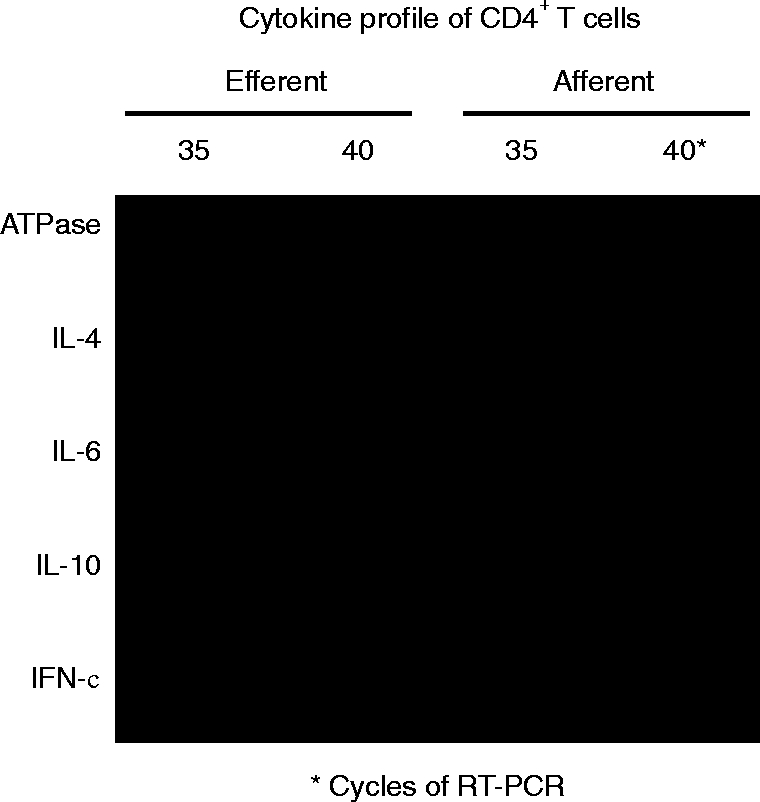
Constitutive cytokine gene transcription by ‘resting’ afferent versus efferent CD4+ T cells. Lymph flow was allowed to settle for 10 days prior to harvesting the cells. 2×10⁷ CD4+ T cells isolated from afferent or efferent lymph by magnetic activated cell sorting (MACS) (>94% purity) were incubated with 0·01mM of α-amatin to block mRNA translation. RNA from 1×10⁷ CD4+ T cells was purified and subjected to reverse transcription–polymerase chain reaction (RT–PCR) using primers for APTase, ovine IL-4, IL-6, IL-10 and IFN-γ. Ten millilitre samples of amplified DNA were slot-blotted onto Hybond-N® using a Biodot® manifold and visualised by Southern hybridization with 32P-labelled cytokine-specific oligonucleotides (internal sequence). The results show that activated CD4+ T cells exhibiting type 1 (IFN-γ) and type 2 (IL-4, IL-6 and IL-10) cytokine expression are concentrated in afferent lymph.
Similar articles
-
Studies on the lymphocytes of sheep. II. Some properties of cells in various compartments of the recirculating lymphocyte pool.Eur J Immunol. 1976 Feb;6(2):121-5. doi: 10.1002/eji.1830060210. Eur J Immunol. 1976. PMID: 1085702
-
Immunological events in the popliteal lymph node of sheep following injection of liver or killed Corynebacterium ovis into an afferent popliteal lymphatic duct.Res Vet Sci. 1977 Jan;22(1):105-12. Res Vet Sci. 1977. PMID: 557223
-
Stochastic regulation of cell migration from the efferent lymph to oxazolone-stimulated skin.J Immunol. 2001 Feb 1;166(3):1517-23. doi: 10.4049/jimmunol.166.3.1517. J Immunol. 2001. PMID: 11160191
-
Lymph node homing of T cells and dendritic cells via afferent lymphatics.Trends Immunol. 2012 Jun;33(6):271-80. doi: 10.1016/j.it.2012.02.007. Epub 2012 Mar 27. Trends Immunol. 2012. PMID: 22459312 Review.
-
[The lymphatic system and its functioning in sheep].Vet Res. 1997 Sep-Oct;28(5):417-38. Vet Res. 1997. PMID: 9410334 Review. French.
Cited by
-
Contribution of Extramedullary Hematopoiesis to Atherosclerosis. The Spleen as a Neglected Hub of Inflammatory Cells.Front Immunol. 2020 Oct 26;11:586527. doi: 10.3389/fimmu.2020.586527. eCollection 2020. Front Immunol. 2020. PMID: 33193412 Free PMC article. Review.
-
Orf Virus-Based Vaccine Vector D1701-V Induces Strong CD8+ T Cell Response against the Transgene but Not against ORFV-Derived Epitopes.Vaccines (Basel). 2020 Jun 10;8(2):295. doi: 10.3390/vaccines8020295. Vaccines (Basel). 2020. PMID: 32531997 Free PMC article.
-
Neutrophil Interactions with the Lymphatic System.Cells. 2021 Aug 17;10(8):2106. doi: 10.3390/cells10082106. Cells. 2021. PMID: 34440875 Free PMC article. Review.
-
In Sickness and in Health: The Immunological Roles of the Lymphatic System.Int J Mol Sci. 2021 Apr 24;22(9):4458. doi: 10.3390/ijms22094458. Int J Mol Sci. 2021. PMID: 33923289 Free PMC article. Review.
-
The beta-chemokine receptor D6 is expressed by lymphatic endothelium and a subset of vascular tumors.Am J Pathol. 2001 Mar;158(3):867-77. doi: 10.1016/s0002-9440(10)64035-7. Am J Pathol. 2001. PMID: 11238036 Free PMC article.
References
-
- Butcher EC, Picker LJ. Lymphocyte homing and homeostasis. Science. 1996;272:60. - PubMed
-
- Hay JB, Cahill RNP. Lymphocyte migration patterns in sheep. In: Hay JB, editor. Animal Models of Immunological Processes. London: Academic Press; 1982. p. 97.
-
- Miyasaka M, Trnka Z. Lymphocyte migration and differentiation in a large animal model: the sheep. Immunol Rev. 1986;91:87. - PubMed
-
- Kimpton WG, Washington EA, Cahill RNP. Lymphocyte recirculation and homing. In: Goddeeris BM, Morrison WI, editors. Cell-Mediated Immunity in Ruminants. Boca Raton: CRC Press; 1994. p. 109.
Publication types
MeSH terms
Substances
Grants and funding
LinkOut - more resources
Full Text Sources
