. 1999 Feb;96(2):176-83.
doi: 10.1046/j.1365-2567.1999.00673.x.

Human eotaxin induces eosinophil extravasation through rat mesenteric venules: role of alpha4 integrins and vascular cell adhesion molecule-1

Affiliations

Human eotaxin induces eosinophil extravasation through rat mesenteric venules: role of alpha4 integrins and vascular cell adhesion molecule-1

K Nagai et al. Immunology. 1999 Feb.

Abstract

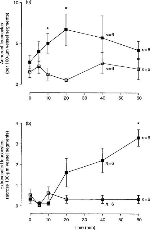
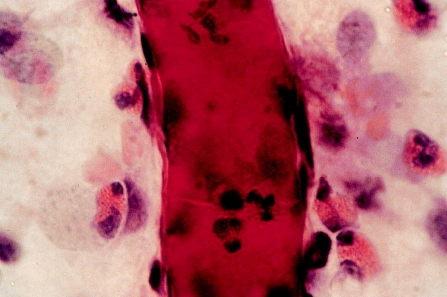
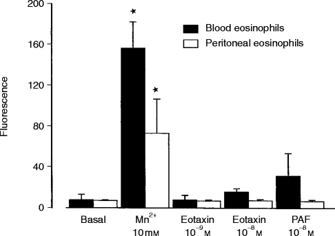
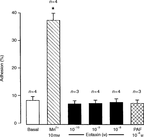
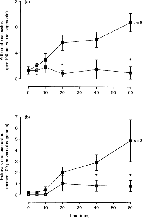
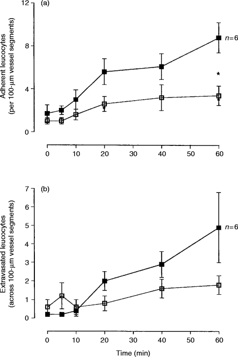
Eotaxin is a potent eosinophil-specific CC-chemokine, which has been shown to play a role in the selective induction of eosinophil accumulation in a number of allergic models of inflammation. Many aspects of the mechanism by which eotaxin induces eosinophil accumulation in vivo remain unresolved. In the present study, we investigated the direct effect of synthetic human eotaxin on leucocyte/endothelial cell interactions within rat mesenteric venules, as quantified by intravital microscopy. Topical eotaxin (30 pmol) induced rapid firm adhesion and extravasation of leucocytes within the rat mesentery, the extravasated leucocytes all being eosinophils, as determined by histological analysis. Whilst eotaxin was unable to stimulate the interaction of rat eosinophils with vascular cell adhesion molecule-1 (VCAM-1) under static conditions in vitro, eotaxin-induced responses in vivo were significantly suppressed by anti-alpha4 integrin and anti-VCAM-1 monoclonal antibodies (mAbs). The anti-alpha4 integrin mAb, HP2/1 (3.5 mg/kg), inhibited the eotaxin-induced firm adhesion and extravasation, 60 min postapplication of the chemokine, by 89% and 84%, respectively. In the same set of experiments, the anti-VCAM-1 mAb, 5F10 (3.5 mg/kg), inhibited leucocyte adhesion and extravasation by 61% and 63%, respectively. These results demonstrate that eotaxin-induced migration of eosinophils through rat mesenteric venules in vivo is dependent on an alpha4 integrin/VCAM-1 adhesion pathway, the significance of which may only be evident under flow conditions and/or following the ligation of other adhesion molecules expressed on eosinophils.


Figures

Figure 1
Figure 1
Effect of topical eotaxin on leucocyte firm adhesion to, and extravasation through, rat mesenteric venules. Graphs show the effect of topical eotaxin (30 pmol, filled square) or Tyrode/BSA (00 μg/ml, open square) on leucocyte (a) firm adhesion and (b) extravasation, over a 60-min time period. Results are presented as mean±SEM for n = 6–7 rats. The data points from n = 6 experiments are indicated on the figure. An asterisk indicates a significant difference between responses detected in eotaxin- and Tyrode/BSA-treated preparations, *P < 0·05.
Figure 2
Figure 2
Histological analysis of an eotaxin-stimulated vessel segment (≈35 μm in diameter) at t = 60 min postapplication of the chemokine. The tissue was stained with haematoxylin and eosin (H&E). The extravasated leucocytes with the grainy red cytoplasm are eosinophils. Magnification ×1000.
Figure 3
Figure 3
Binding of immunoglobulin–vascular cell adhesion molecule-1 (immunoglobulin–VCAM-1) to rat eosinophils as determined by indirect immunostaining and fluorescence-activated cell sorter (FACS) analysis. Whole blood or purified rat peritoneal eosinophils were co-incubated with phosphate-buffered saline (basal), Mn2+, eotaxin or platelet-activating factor (PAF) and immunoglobulin–VCAM-1 (or the control chimaera immunoglobulin–lymphocyte function-associated antigen-3) for 30 min, washed and then incubated with a fluorescein isothiocyanate-labelled mouse antihuman IgG, as described in the Materials and methods. Binding of immunoglobulin–VCAM-1 to eosinophils was quantified by measuring the geometric mean fluorescence intensity of eosinophils in samples incubated with immunoglobulin–VCAM-1, corrected for the fluorescence intensity detected in corresponding control samples treated with immunoglobulin-LFA-3. Results show mean±SEM for n = 3 experiments. An asterisk indicates a significant difference from basal levels of fluorescence, P < 0·05.
Figure 4
Figure 4
Adhesion of rat eosinophils to recombinant soluble vascular cell adhesion molecule-1 (rsVCAM-1)-coated plates. Purified rat peritoneal eosinophils were incubated with stimuli in 96-well plates coated with rsVCAM-1, as described in the Materials and methods. Results are presented as mean±SEM of percentage adhesion from n = 3–4 experiments. An asterisk indicates a significant difference from basal levels of adhesion, *P < 0·05.
Figure 5
Figure 5
Effect of the anti-α4 integrin monoclonal antibody (mAb), HP2/1, on eotaxin-induced leucocyte responses within rat mesenteric venules. Rats were treated with MOPC-21 (filled square) (control mouse IgG, 3·5 mg/kg i.v., n = 7 unless indicated on the figure) or the anti-α4 integrin mAb, HP2/1 (open square) (3·5 mg/kg, i.v., n = 4), prior to the topical application of human eotaxin (30 pmol). The graphs show the effects of HP2/1 on leucocyte (a) firm adhesion and (b) extravasation, over a 60-min time period. Results are presented as mean±SEM for n = 4–7 rats as detailed above and on the figures. An asterisk indicates a significant difference between responses detected in MOPC-21- and HP2/1-treated rats, *P < 0·05.
Figure 6
Figure 6
Effect of the anti-vascular cell adhesion molecule-1 (VCAM-1) monoclonal antibody (mAb), 5F10, on eotaxin-induced leucocyte responses within rat mesenteric venules. Rats were treated with MOPC-21 (filled square) (control mouse IgG, 3·5 mg/kg i.v., n = 7 unless indicated on the figure) or the anti-VCAM-1 mAb 5F10 (open square) (3·5 mg/kg, i.v., n = 5), prior to the topical application of human eotaxin (30 pmol). The graphs show the effects of 5F10 on leucocyte (a) firm adhesion and (b) extravasation, over a 60-min time period. Results are presented as mean±SEM for n = 5–7 rats, as indicated above and on the figures. An asterisk indicates a significant difference between responses detected in MOPC-21- and 5F10-treated rats, *P < 0·05.

Similar articles

Cited by

References

    1. Griffiths-Johnson DA, Collins PD, Rossi AG, Jose PJ, Williams TJ. The chemokine, eotaxin, activates guinea-pig eosinophils in vitro, and causes their accumulation into the lung in vivo. Biochem Biophys Res Commun. 1993;197:1167. - PubMed
    1. Jose PJ, Griffiths-Johnson DA, Collins PD, et al. Eotaxin: a potent eosinophil chemoattractant cytokine detected in a guinea-pig model of allergic airways inflammation. J Exp Med. 1994;179:881. - PMC - PubMed
    1. Collins PD, Marleau S, Griffiths-Johnson DA, Jose PJ, Williams TJ. Co-operation between interleukin-5 and the chemokine eotaxin, to induce eosinophil accumulation in vivo. J Exp Med. 1995;182:1169. - PMC - PubMed
    1. Humbles AA, Conroy DM, Marleau S, et al. Kinetics of eotaxin generation and its relationship to eosinophil accumulation in allergic airways disease: analysis in a guinea pig model in vivo. J Exp Med. 1997;186:601. - PMC - PubMed
    1. Palframan RT, Collins PD, Williams TJ, Rankin SM. Eotaxin induces a rapid release of eosinophils and their progenitors from the bone marrow. Blood. 1998;91:2240. - PubMed

Publication types

MeSH terms

LinkOut - more resources