

Effect of CD8+ T-cell depletion on bronchial hyper-responsiveness and inflammation in sensitized and allergen-exposed Brown-Norway rats
- PMID: 10233723
- PMCID: PMC2326765
- DOI: 10.1046/j.1365-2567.1999.00699.x
Effect of CD8+ T-cell depletion on bronchial hyper-responsiveness and inflammation in sensitized and allergen-exposed Brown-Norway rats
Abstract
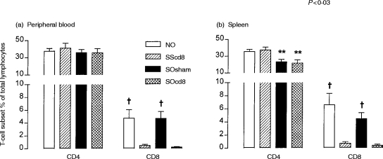
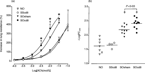
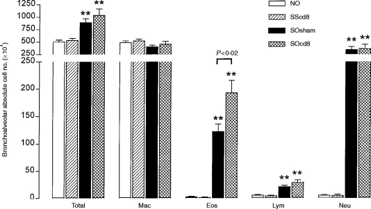
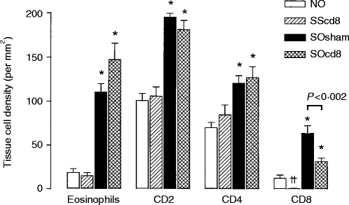
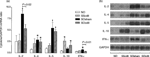
We examined the role of CD8+ T cells in a Brown-Norway rat model of asthma, using a monoclonal antibody to deplete CD8+ T cells. Ovalbumin (OA)-sensitized animals were given anti-CD8 antibody (0.5 mg/rat) intravenously 1 week prior to exposure to 1% OA aerosol and were studied 18-24 hr after aerosol exposure. Following administration of anti-CD8 antibody, CD8+ cells were reduced to <1% of total lymphocytes in whole blood and in spleen. In sensitized animals, OA exposure induced bronchial hyper-responsiveness (BHR), accumulation of eosinophils, lymphocytes and neutrophils in bronchoalveolar lavage (BAL) fluid, and also an increase in tissue eosinophils and CD2+, CD4+ and CD8+ T cells in airways. Anti-CD8 antibody caused a further increase in allergen-induced BHR (P<0.03, compared with sham-treated animals), together with a significant increase in eosinophil number in BAL fluid (P<0.05). While CD2+ and CD4+ T cells in airways were not affected by anti-CD8 treatment, the level of CD8+ T cells was significantly reduced in sensitized, saline-exposed animals (P<0.04, compared with sham-treated rats), and sensitized and OA-challenged rats (P<0.002, compared with sham-treated rats). Using reverse transcription-polymerase chain reaction, an increase of T helper (Th)2 cytokine [interleukin (IL)-4 and IL-5], and also of Th1 cytokine [interferon-gamma (IFN-gamma) and IL-2], mRNA in the lung of sensitized and OA-exposed animals was found; after CD8+ T-cell depletion, Th1 cytokine expression was significantly reduced (P<0.02), while Th2 cytokine expression was unchanged. CD8+ T cells have a protective role in allergen-induced BHR and eosinophilic inflammation, probably through activation of the Th1 cytokine response.
Figures
Similar articles
-
Inhibitory effects of endogenous and exogenous interferon-gamma on bronchial hyperresponsiveness, allergic inflammation and T-helper 2 cytokines in Brown-Norway rats.Immunology. 1999 Oct;98(2):280-8. doi: 10.1046/j.1365-2567.1999.00870.x. Immunology. 1999. PMID: 10540228 Free PMC article.
-
Differential regulation of cytokine expression after allergen exposure of sensitized rats by cyclosporin A and corticosteroids: relationship to bronchial hyperresponsiveness.J Allergy Clin Immunol. 1999 Sep;104(3 Pt 1):644-52. doi: 10.1016/s0091-6749(99)70337-4. J Allergy Clin Immunol. 1999. PMID: 10482841
-
T-cells subsets and activation in bronchial mucosa of sensitized Brown-Norway rats after single allergen exposure.Immunology. 1995 Aug;85(4):591-7. Immunology. 1995. PMID: 7558154 Free PMC article.
-
CD8+ T lymphocytes and leukotriene B4: novel interactions in the persistence and progression of asthma.J Allergy Clin Immunol. 2006 Mar;117(3):577-82. doi: 10.1016/j.jaci.2005.12.1340. J Allergy Clin Immunol. 2006. PMID: 16522456 Review.
-
Essential role of T lymphocytes in the development of allergen-driven airway hyperresponsiveness.Allergy Asthma Proc. 1998 Nov-Dec;19(6):365-9. doi: 10.2500/108854198778612744. Allergy Asthma Proc. 1998. PMID: 9876776 Review.
Cited by
-
Interferon-gamma secretion of peripheral blood CD8+ T lymphocytes in patients with bronchial asthma: in vitro stimulus determines cytokine production.Clin Exp Immunol. 2001 Nov;126(2):199-205. doi: 10.1046/j.1365-2249.2001.01666.x. Clin Exp Immunol. 2001. PMID: 11703361 Free PMC article.
-
Airway hyperresponsiveness in the absence of CD4+ T cells after primary but not secondary challenge.Am J Respir Cell Mol Biol. 2005 Jul;33(1):89-96. doi: 10.1165/rcmb.2004-0414OC. Epub 2005 Apr 21. Am J Respir Cell Mol Biol. 2005. PMID: 15845865 Free PMC article.
-
Complement mediators: key regulators of airway tissue remodeling in asthma.J Transl Med. 2015 Aug 20;13:272. doi: 10.1186/s12967-015-0565-2. J Transl Med. 2015. PMID: 26289385 Free PMC article. Review.
-
Role of CD8-positive cells in radioimmunotherapy utilizing (177)Lu-mAbs in an immunocompetent rat colon carcinoma model.EJNMMI Res. 2015 Feb 12;5:3. doi: 10.1186/s13550-014-0079-6. eCollection 2015. EJNMMI Res. 2015. PMID: 25853009 Free PMC article.
-
A novel transcription factor inhibitor, SP100030, inhibits cytokine gene expression, but not airway eosinophilia or hyperresponsiveness in sensitized and allergen-exposed rat.Br J Pharmacol. 2001 Nov;134(5):1029-36. doi: 10.1038/sj.bjp.0704344. Br J Pharmacol. 2001. PMID: 11682451 Free PMC article.
References
-
- Azzawi M, Bradley B, Jeffery PK, et al. Identification of activated T lymphocytes and eosinophils in bronchial biopsies in stable atopic asthma. Am Rev Respir Dis. 1990;142:1407. - PubMed
-
- Robinson DS, Hamid Q, Ying S, et al. Predominant Th2-like bronchoalveolar T-lymphocyte populations in atopic asthma. N Engl J Med. 1992;326:298. - PubMed
-
- Romagniani S. Regulation and deregulation of human IgE synthesis. Immunol Today. 1990;11:316. - PubMed
-
- Sanderson CJ. Interleukin-5, eosinophils and disease. Blood. 1992;79:3101. - PubMed
MeSH terms
Substances
LinkOut - more resources
Full Text Sources
Medical
Research Materials
