. 1999 Mar;96(3):440-6.
doi: 10.1046/j.1365-2567.1999.00700.x.

Importance of intrathymic mixed chimerism for the maintenance of skin allograft tolerance across fully allogeneic antigens in mice

Affiliations

Importance of intrathymic mixed chimerism for the maintenance of skin allograft tolerance across fully allogeneic antigens in mice

M Eto et al. Immunology. 1999 Mar.

Abstract

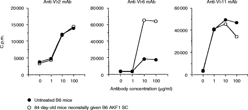
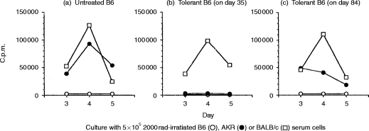
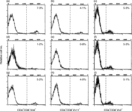
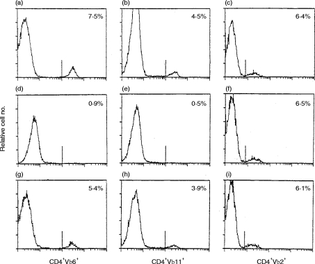
In B6 (H-2b) mice that had been given, neonatally, 1x10⁸ B6AKF1 spleen cells intraperitoneally (i.p.), only a moderate prolongation of donor (AKR:H-2k) skin graft survival was observed. In such B6 mice, no mixed lymphocyte reaction (MLR) to AKR could be detected on day 35 (35 days after birth), but it was clearly evident on day 84. Similarly, neither Vbeta6+ (reactive to MTV-7-encoded antigens) nor Vbeta11+ (reactive to I-E+MTV-derived superantigens) T cells were detected on day 35, but both were clearly evident on day 84 in both the thymus and the lymph nodes, thus indicating the breakdown of intrathymic mixed chimerism at the antigen-presenting cell level. Furthermore, by day 84, all skin grafts from AKR had already been rejected in such B6 mice. In the periphery, however, Vbeta6+, but not Vbeta11+, T cells were clonally anergic on day 84, based on a stimulation assay with anti-T-cell receptor (TCR) monoclonal antibody (mAb), thus suggesting that tolerance to some antigens, but not to others, may be induced by the clonal anergy in fully allogeneic combinations, and that the clonal anergic state may be masked by other proliferative responses. These results therefore indicate the importance of intrathymic mixed chimerism (central tolerance) and the limitations of clonal anergy (peripheral tolerance) in maintaining tolerance across fully allogeneic antigen barriers.


Figures

Figure 4
Figure 4
T cells using T-cell receptor (TCR) Vβ6, but not Vβ11, were clonally anergic in the late stage of tolerance. For a monoclonal antibody (mAb)-induced proliferation assay, the purified mAbs 44-22-1, KT11, or B20.6 (specific for TCR Vβ6, Vβ11, or Vβ2, respectively) were diluted to the indicated concentrations in phosphate-buffered saline (PBS) and 30 μl was added per round-bottomed microtitre well. The plates were incubated for 4 to 6 hr at 37° and then washed three times with PBS before use. Lymph node cells (2×10⁵ in 200 μl of RPMI medium) were added to each well, cultured for an optimal 48 hr, pulsed for the last 6 hr with 1 μCi 3H-thymidine and then harvested. The lymph node cells were obtained from both the untreated B6 mice and the 84-day-old B6 mice that had been given B6AKF1 SC neonatally. All values represent the arithmetic means of triplicate cultures. The standard deviation (SD) was generally less than 10% of the mean.
Figure 1
Figure 1
B6 mice that had been given B6AKF1 spleen cells (SC) neonatally were tolerant to AKR on day 35, but not on day 84, in a mixed lymphocyte reaction (MLR). SC (5×10⁵) from the untreated B6 mice (a), the 5-week-old B6 mice that had been made tolerant, neonatally, with B6AKF1 SC (day 35) (b), or the 12-week-old B6 mice that had been made tolerant, neonatally, with B6AKF1 SC (day 84) (c), were cultured with 5×10⁵ 2000 rad-irradiated B6, AKR or BALB/c SC, as indicated on the Figure. The responder cells and stimulator cells were incubated for 3, 4 or 5 days, and were pulsed for the last 12 hr on each day with 3H-thymidine (1 μCi/well) and then harvested.
Figure 2
Figure 2
Neither Vβ6+ nor Vβ11+ T cells were detected on day 35, but both were clearly evident on day 84 in the thymus of the recipient B6 mice. The whole thymocytes were triple stained with phycoerythrin (PE)-conjugated anti-CD4 monoclonal antibody (mAb), DC-conjugated anti-CD8 mAb and the mAbs 44-2-1, KT11 or B20.6 (specific for T-cell receptor (TCR) Vβ6, Vβ11 or Vβ2, respectively) followed by fluorescein isothiocyanate (FITC)-conjugated goat antirat immunoglobulin G (IgG) mAb. CD4+CD8 or CD4CD8+ single-positive thymocytes were selected by gating based on CD4 (PE) fluorescence and CD8 (DC) fluorescence. Only the data for CD4+CD8 thymocytes stained with individual anti-TCR mAb were illustrated (representing 1–2×10⁴ viable cells). The percentage of T cells expressing the individual TCR, Vβ, relative to the total number of CD4+CD8 single-positive thymocytes, was given. Thymocytes were obtained from: untreated B6 mice (a), (b) and (c); the 35-day-old B6 mice that had been given B6AKF1 SC neonatally (d), (e) and (f); and the 84-day-old B6 mice that had been given B6AKF1 SC neonatally (g), (h) and (i). The data represent one of the three animals tested. The other two animals showed similar staining patterns in each panel.
Figure 3
Figure 3
Neither Vβ6+ nor Vβ11+ T cells were detected on day 35, but both of them were clearly evident on day 84 in the lymph node cells of the recipient B6 mice. The lymph node cells were double stained with phycoerythrin (PE)-conjugated anti-CD4 monoclonal antibody (mAb) and the mAbs 44-22-1, KT11, or B20.6 (specific for T-cell receptors (TCR) Vβ6, Vβ11, or Vβ2, respectively) followed by fluorescein isothiocyanate (FITC)-conjugated goat antirat immunoglobulin G (IgG) mAb. Only the data for CD4+ T cells stained with individual anti-TCR mAb were illustrated (representing 1–2×10⁴ viable cells). The percentage of T cells expressing the individual TCR, Vβ, relative to the total number of the CD4+ T cells, was given. The lymph node cells were obtained from: untreated B6 mice (a), (b) and (c); the 35-day-old B6 mice that had been given B6AKF1 spleen cells (SC) neonatally (d), (e) and (f); and the 84-day-old B6 mice that had been given B6AKF1 SC neonatally (g), (h) and (i). The data represent one of the three animals tested. The other two animals showed similar staining patterns in each panel.

Similar articles

Cited by

References

    1. Good RA, Martinez C, Gabrielsen AE. Progress toward transplantation of tissues in man. Adv Pediatr. 1964;13:93. - PubMed
    1. Billingham RE, Brent L, Medawar PB. ‘Actively aquired tolerance’ of foreign cells. Nature. 1953;172:603. - PubMed
    1. Morrissey PJ, Sharrow SO, Kohno Y, Berzofsky JA, Singer A. Correlation of intrathymic tolerance with intrathymic chimerism in neonatally tolerized mice. Transplantation. 1985;40:68. - PubMed
    1. Ildstad ST, Sachs DH. Reconstitution with syngeneic plus allogeneic or xenogeneic bone marrow leads to specific acceptance of skin allografts or xenografts. Nature. 1984;307:168. - PubMed
    1. Sharabi Y, Sachs DH. Mixed chimerism and permanent specific transplantation tolerance induced by a nonlethal preparative regimen. J Exp Med. 1989;169:493. - PMC - PubMed

Publication types

Substances

LinkOut - more resources