Review
. 1999 Apr;154(4):965-73.
doi: 10.1016/S0002-9440(10)65346-1.

Molecular evolution of the metaplasia-dysplasia-adenocarcinoma sequence in the esophagus

Affiliations
Review

Molecular evolution of the metaplasia-dysplasia-adenocarcinoma sequence in the esophagus

J A Jankowski et al. Am J Pathol. 1999 Apr.

Abstract

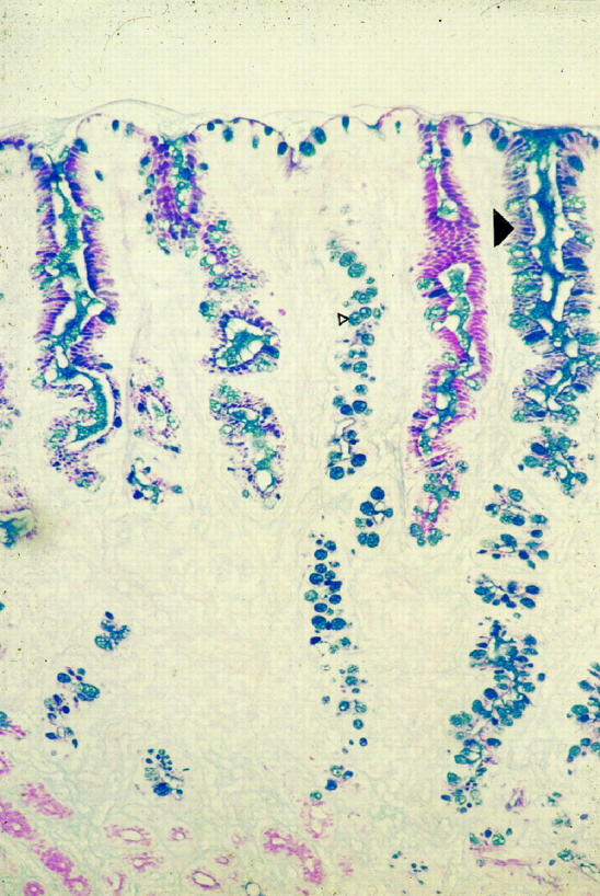
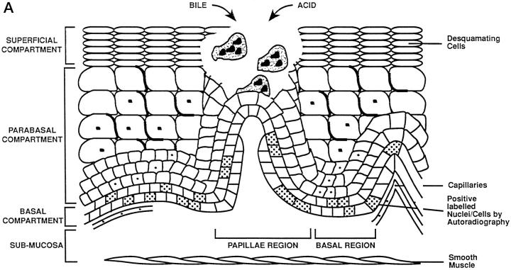
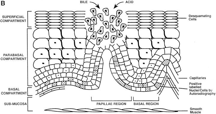
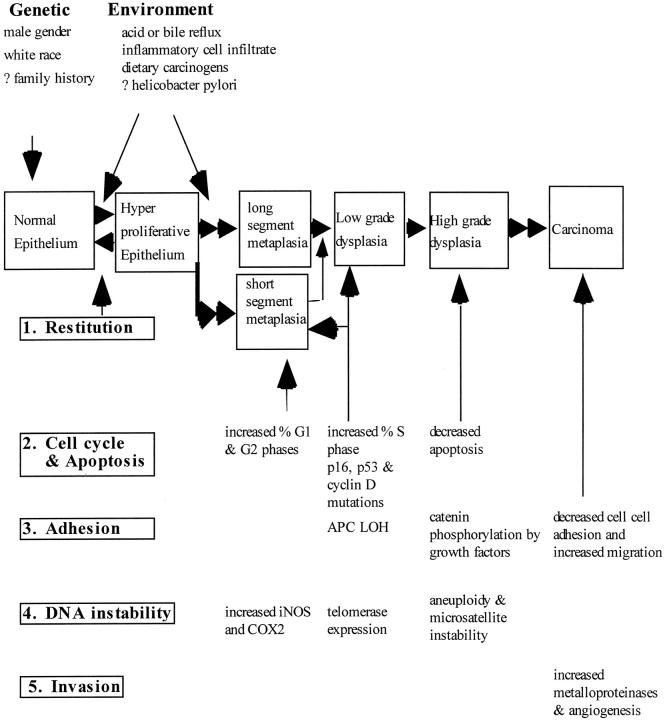
The incidence of adenocarcinoma of the esophagus has been increasing in developing countries over the last three decades and probably reflects a genuine increase in the incidence of its recognized precursor lesion, Barrett's metaplasia. Despite advances in multimodality therapy, the prognosis for invasive esophageal adenocarcinoma is poor. An improved understanding of the molecular biology of this disease may allow improved diagnosis, therapy, and prognosis. We focus on recent developments in the molecular and cell biology of Barrett's metaplasia, a heterogeneous lesion affecting the transitional zone of the gastro-esophageal junction whose associated molecular alterations may vary both in nature and temporally. Early premalignant clones produce biological and genetic heterogeneity as seen by multiple p53 mutations, p16 mutations, aneuploidy, and abnormal methylation resulting in stepwise changes in differentiation, proliferation, and apoptosis, allowing disease progression under selective pressure. Abnormalities in expression of growth factors of the epidermal growth factor family and cell adhesion molecules, especially cadherin/catenin complexes, may occur early in invasion. Exploitation of these molecular events may lead to a more appropriate diagnosis and understanding of these lesions in the future.


Figures

Figure 1.
Figure 1.
Photomicrograph of intestinal metaplasia in Barrett’s esophagus stained with Alcian blue/periodic acid-Schiff (mucins). Barrett’s esophagus is composed of columnar lined mucus-secreting cells and a proportion of the glands will be composed of goblet cells (small arrowhead). Alcian blue diastase periodic acid-Schiff staining indicates the heterogeneity of mucin phenotypes in esophageal cells: blue (basic), red (neutral), and purple (mixed) mucins (large arrowhead). Original magnification, ×250.
Figure 2.
Figure 2.
Schematic representation of adaptation during Barrett’s mucosa formation. The three compartments of the esophageal epithelium are represented on the diagrams. The bottom of each diagram shows basal compartment containing both stem cells (speckled nucleus) and proliferating cells; the middle, parabasal compartment containing proliferating cells; and the top, superficial compartment containing only mature differentiated cells. The uninflamed mucosa (right) is flat, whereas the inflamed mucosa (left) has invaginations of the basal layer, termed papillae. A: Damage to the esophageal differentiated cells in the superficial and parabasal compartments of the esophagus. B: Damage to a deeper compartment involving the squamous epithelial stem cells in the basal compartment of the papillae. C: Generation of clones with a mucin-secreting lineage resistant to acid/bile.
Figure 2.
Figure 2.
Schematic representation of adaptation during Barrett’s mucosa formation. The three compartments of the esophageal epithelium are represented on the diagrams. The bottom of each diagram shows basal compartment containing both stem cells (speckled nucleus) and proliferating cells; the middle, parabasal compartment containing proliferating cells; and the top, superficial compartment containing only mature differentiated cells. The uninflamed mucosa (right) is flat, whereas the inflamed mucosa (left) has invaginations of the basal layer, termed papillae. A: Damage to the esophageal differentiated cells in the superficial and parabasal compartments of the esophagus. B: Damage to a deeper compartment involving the squamous epithelial stem cells in the basal compartment of the papillae. C: Generation of clones with a mucin-secreting lineage resistant to acid/bile.
Figure 3.
Figure 3.
Schematic representation of the key molecular events in Barrett’s dysplasia, the metaplasia-dysplasia-adenocarcinoma sequence (MCS). Acid and bile cause acute damage to the esophagus, which is rapidly healed by restitution or cellular replication (stages 1 and 2). In 10% of cases chronic damage to the epithelial stem cells allows rapid clonal replacement by lineages with a growth advantage containing p53 mutations (stage 2). The formation of each type of Barrett’s metaplasia is dependent on the stem cell from which it arises as well as the nature of the mucosal microenvironment. Appearance of dysplasia is associated in part with loss of heterozygosity of APC or alterations in the catenins (stage 3). In 1 in 100 cases, aneuploidy and errors in DNA repair represent final pathways which disrupt invasion suppressor genes (stages 4 and 5). The transition from high grade dysplasia to invasive cancer is rapid in all cases.

References

    1. Spechler SJ: Barrett’s esophagus. Sem Oncol 1994, 21:431-437 - PubMed
    1. Winter C, Jr, Spurling TJ, Chobabian SJ, Curtis DJ, Esposito RL, Hacker JF, III, Johnston DA, Cruess DF, Cotelingam JD, Gurney MS, Cattau EL, Jr: Barrett’s esophagus: a prevalent, occult complication of gastroesophageal reflux disease. Gastroenterology 1987, 92:118-124 - PubMed
    1. Prach AT, MacDonald TA, Hopwood DA, Johnston DA: Increasing incidence of Barrett’s oesophagus: education, enthusiasm or epidemiology. Lancet 1997, 350:933 - PubMed
    1. Beynon J, Pye JK, Howell P, Nehra D: Assessment of combined bile acid and pH profiles using an automated sampling device in gastro-oesophageal reflux disease. Br J Surg 1998, 85:134-137 - PubMed
    1. Romero Y, Cameron AJ, Locke GR, Schaid DJ, Melton LJ, Slexak J: Familial gastroesophageal reflux: association with Barrett’s esophagus and adenocarcinoma. Gastroenterology 1997, 110:456 - PubMed

Publication types

MeSH terms

Substances