. 1999 Apr;154(4):1057-65.
doi: 10.1016/S0002-9440(10)65358-8.

Mechanisms of enhanced lung injury during sepsis

Affiliations

Mechanisms of enhanced lung injury during sepsis

B J Czermak et al. Am J Pathol. 1999 Apr.

Abstract

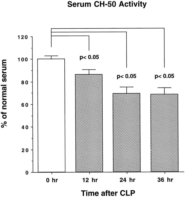
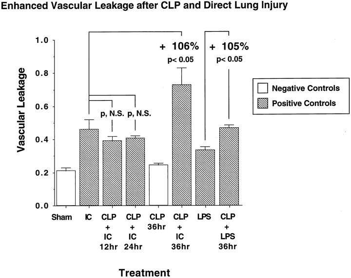
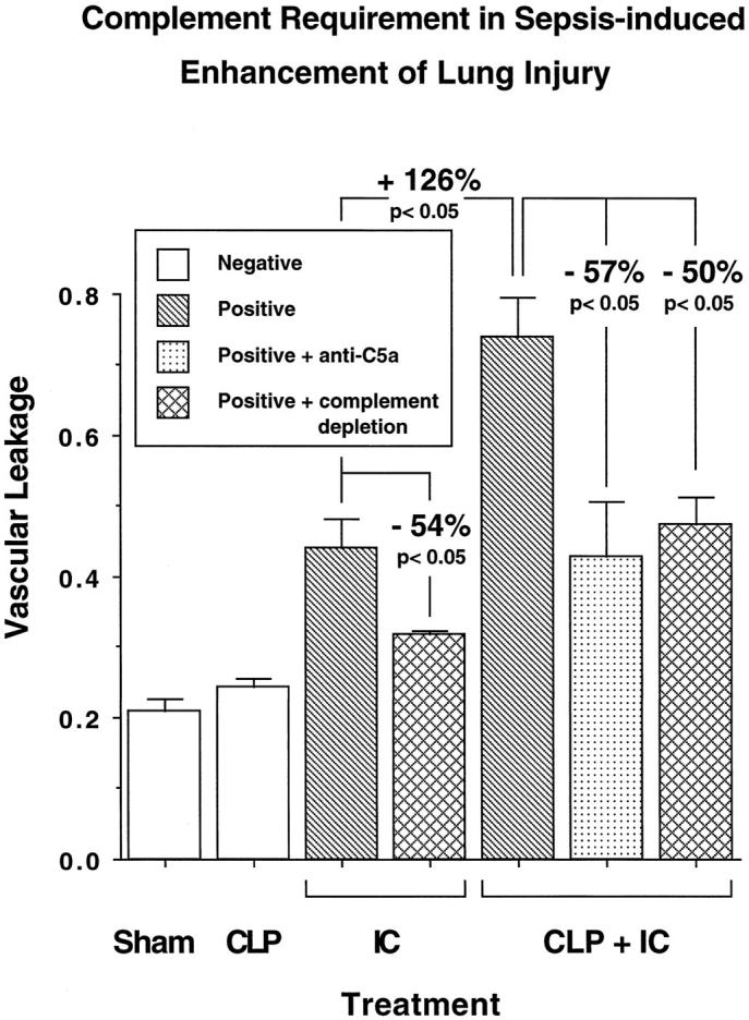
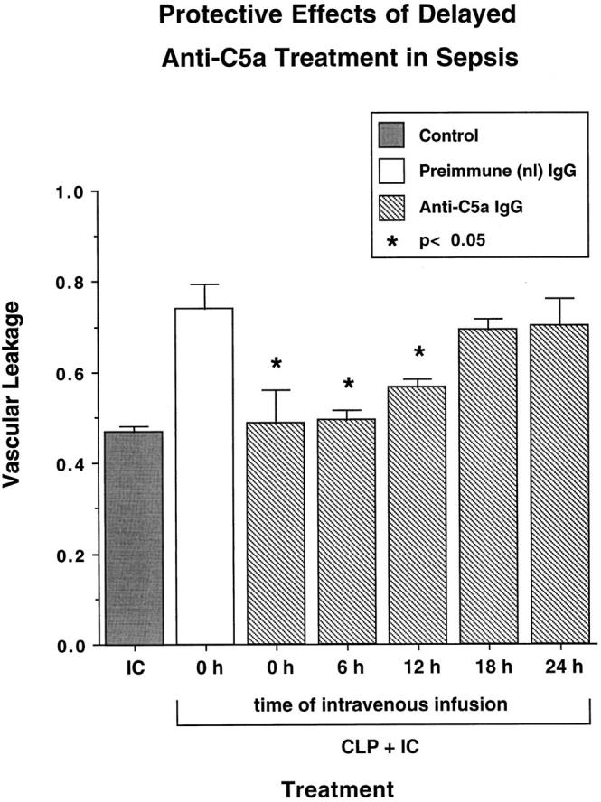
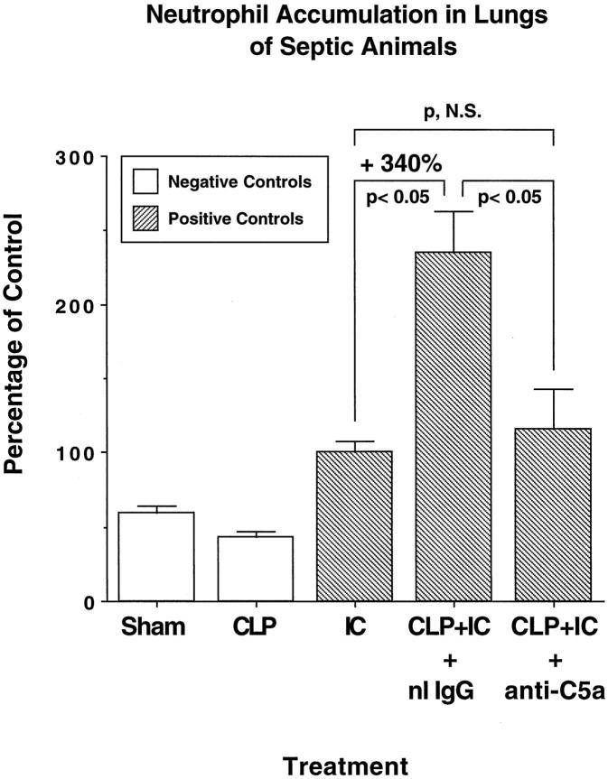
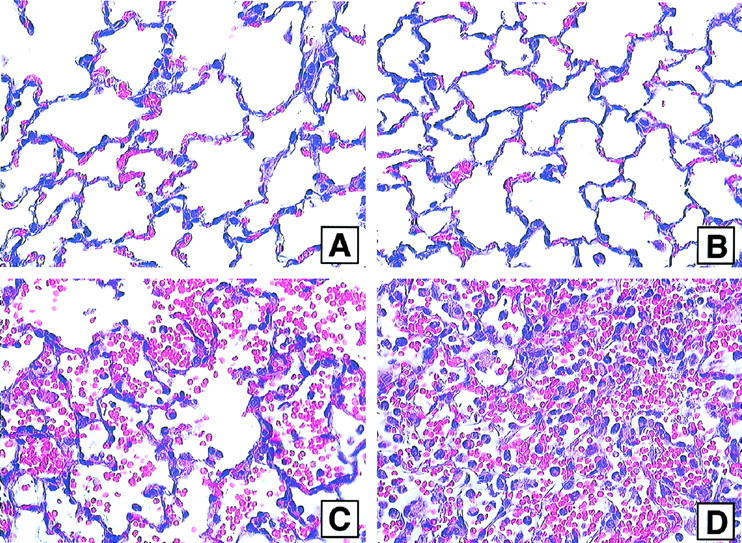
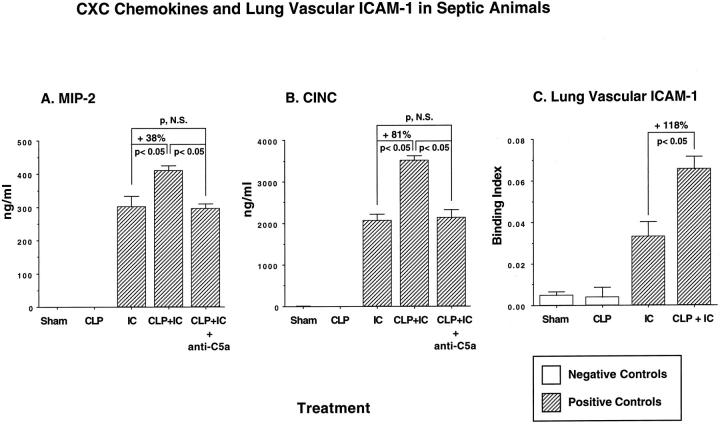
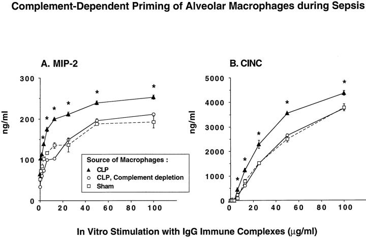
A major complication in sepsis is progressively impaired lung function and susceptibility to intrapulmonary infection. Why sepsis predisposes the lung to injury is not clear. In the current studies, rats were rendered septic by cecal ligation/puncture and evaluated for increased susceptibility to injury after a direct pulmonary insult (deposition of IgG immune complexes or airway instillation of lipopolysaccharide). By itself, cecal ligation/puncture did not produce evidence of lung injury. However, after a direct pulmonary insult, lung injury in septic animals was significantly enhanced. Enhanced lung injury was associated with increased accumulation of neutrophils in lung, enhanced production of CXC chemokines (but not tumor necrosis factor-alpha) in bronchoalveolar lavage fluids, and increased expression of lung vascular intercellular adhesion molecule-1 (ICAM-1). Complement depletion or treatment with anti-C5a abolished all evidence of enhanced lung injury in septic animals. When stimulated in vitro, bronchoalveolar lavage macrophages from septic animals had greatly enhanced CXC chemokine responses as compared with macrophages from sham-operated animals or from septic animals that had been complement depleted. These data indicate that the septic state causes priming of lung macrophages and suggest that enhanced lung injury in the septic state is complement dependent and related to increased production of CXC chemokines.


Figures

Figure 1.
Figure 1.
Serum complement hemolytic activity (CH50) of animals undergoing CLP-induced sepsis. Blood samples were collected via an intra-arterial catheter. Sensitized sheep erythrocytes were exposed to various dilutions of serum samples, and hemolysis was detected with a spectrophotometer at 541 nm. For easier comparability, absolute values of the measured absorbance were calculated to the percentage of normal serum (100%); n = 4. See text for details.
Figure 2.
Figure 2.
Enhanced lung vascular leakage 12 to 36 hours after CLP in the presence or absence of a direct lung insult (intrapulmonary deposition of IgG immune complexes (IC) or intratracheal instillation of LPS). The lung vascular permeability index, referred to in this and in subsequent figures as vascular leakage, was assessed by measuring the extravasation of intravenously administered 125I-labeled BSA for 4 hours in the immune complex model and 6 hours in the LPS model. Open bars represent negative control groups; hatched bars represent positive controls. Calculations of percent change were determined by first subtracting the negative control values from the positive control groups. See text for details. For this and all subsequent groups, n ≥ 5.
Figure 3.
Figure 3.
Complement requirements for enhanced lung injury in septic animals. Anti-C5a was administered intravenously at the time of CLP (36 hours before IgG immune complex-induced lung injury (IC)). Positive and negative control groups received preimmune IgG instead. Complement depletion was achieved by repetitive intraperitoneal injections of CVF before CLP.
Figure 4.
Figure 4.
Protective effects of delayed anti-C5a treatment until after induction of sepsis. Animals were subjected to CLP, followed by a single intravenous administration of 300 μg of anti-C5a after indicated intervals. Control animals received 300 μg of preimmune goat IgG at the time of CLP. At 36 hours after CLP a direct pulmonary insult by intra-alveolar IgG immune complex (IC) deposition was induced, and lung vascular permeability was assessed 4 hours later by detection of extravasation of albumin (vascular leakage). In one control group, immune complexes were instilled intratracheally in the absence of sepsis. Statistical differences shown are in comparison with the septic group treated with immune complexes in the presence of preimmune IgG.
Figure 5.
Figure 5.
Neutrophil content in BAL fluids. BAL samples were centrifuged, and pellets were resuspended for differential cell counts. For matters of simplicity, data are expressed as percentage of values obtained in nonseptic positive controls. Absolute values are described in the text. As indicated, measurements were made 4 hours after initiation of IC reactions and 36 hours after initiation of CLP. When used, goat anti-C5a or preimmune goat IgG (each at 300 μg) was injected intravenously.
Figure 6.
Figure 6.
Lung sections obtained from animals undergoing sham surgery (A), CLP alone (B) or intra-alveolar deposition of IgG immune complexes alone (C) or in combination with CLP induced 36 hours earlier (D). H&E; magnification, ×40.
Figure 7.
Figure 7.
BAL content of CXC chemokines (A and B) and lung vascular ICAM-1 (C) 36 hours after CLP-induced sepsis in the presence or absence of an intrapulmonary insult (deposition of IC). Quantitation of chemokines was achieved using sandwich ELISA techniques; up-regulation of lung vascular ICAM-1 was determined by binding of 125I-labeled anti-ICAM-1 as described in the text. Sham-operated animals underwent surgery without CLP.
Figure 8.
Figure 8.
Dose responses for in vitro chemokine generation by rat alveolar macrophages obtained from sham-operated animals or 36 hours after CLP in the presence or absence of complement depletion. A total of 10 macrophages were plated in tissue culture wells followed by addition of a range of concentrations of immune complexes. Supernatant fluids were collected 4 hours later for ELISA analysis of MIP-2 and CINC. Results are shown using macrophages from CLP animals, from CLP animals with complement depletion, and from sham-operated animals. *Statistical difference between the CLP group and the two accompanying groups at the same dose of stimulus.

References

    1. Young LS: Gram-negative sepsis. Mandell GL Douglas SD Bennett JC eds. Principles and Practice of Infectious Diseases. 1979, :pp 571-608 John Wiley and Sons, New York
    1. Bone RC: The sepsis syndrome: definition and general approach to management. Clin Chest Med 1996, 17:175-181 - PubMed
    1. Kaplan RL, Sahn SA, Petty TL: Incidence and outcome of respiratory distress syndrome in gram-negative sepsis. Arch Intern Med 1979, 139:867-869 - PubMed
    1. Moore FA, Moore EE, Read RA: Postinjury multiple organ failure: role of extrathoracic injury and sepsis in adult respiratory distress syndrome. New Horizons 1993, 1:538-549 - PubMed
    1. Polk HC, Shields CL: Remote organ failure: a valid sign of occult intra-abdominal infection. Surgery 1977, 81:310-331 - PubMed

Publication types

MeSH terms