. 1999 Apr;154(4):1105-14.
doi: 10.1016/S0002-9440(10)65363-1.

Human trophoblast invasion and spiral artery transformation: the role of nitric oxide

Affiliations

Human trophoblast invasion and spiral artery transformation: the role of nitric oxide

F Lyall et al. Am J Pathol. 1999 Apr.

Abstract

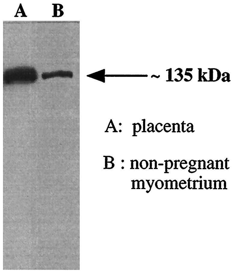
During early human pregnancy extravillous cytotrophoblasts invade the uterus and also migrate up the spiral arteries, transforming them into large vessels of low resistance. Failure of transformation has been described in pre-eclampsia, fetal growth restriction, and miscarriage. Recent evidence suggests that some maternal vessels undergo structural changes without interaction with cytotrophoblasts. The possibility arises that local vasoactive mediators such as nitric oxide result in spiral artery dilatation before their invasion. In support of this, a recent histological study in the guinea pig suggested that cytotrophoblasts expressed nitric oxide synthase (NOS) as they surrounded vessels. This study tested the hypothesis that invading cytotrophoblasts express NOS and therefore have the potential to induce vasodilatation by releasing nitric oxide. The expression of NOS on extravillous cytotrophoblasts was studied in placental bed biopsies, obtained, using a transcervical sampling technique, from normal human pregnancies between 8 to 19 weeks of gestation and in the third trimester. Whereas eNOS was expressed by syncytiotrophoblast, neither eNOS or iNOS was expressed by extravillous cytotrophoblasts at any time during invasion. The mechanisms controlling spiral artery transformation are pivotal to understanding normal and abnormal placentation. These results suggest that trophoblast-derived nitric oxide is unlikely to contribute to spiral artery dilatation.


Figures

Figure 1.
Figure 1.
Western blot showing an eNOS immunoreactive band at approximately 135 kd in a membrane preparation of term placental villous tissue (A) and a membrane preparation of nonpregnant myometrium (B). Forty-μg membrane protein was loaded in each lane.
Figure 2.
Figure 2.
Western blot with the iNOS antibodies. A in both blots is ADP-sepharose extracted proteins from unstimulated A549 cells and no band was evident. B in both blots is the cytokine cocktail stimulated cells (100 μmol/L lipopolysaccaride, 10 ng/ml interferon-γ, 10 ng/ml tumor necrosis factor-α, 10 ng/ml interleukin-1β). An iNOS immunoreactive band at approximately 130 kd is clearly identifiable with both antibodies. Twenty μl of reaction mix was loaded in each lane.
Figure 3.
Figure 3.
Expression of cytokeratin (A, E, G) and eNOS (B, C, D, F, H) on placental cytotrophoblast columns/islands at 8 weeks of gestation (A and B, cell island), 11 weeks of gestation (C, cytotrophoblast cell column), 14 weeks of gestation (D, cytotrophoblast cell column), 16 weeks of gestation (E and F, cytotrophoblast cell column), and 19 weeks of gestation (cytotrophoblast cell island, G and H). Cytotrophoblast cell islands are indicated by double arrows and cell columns by single arrows. Cytokeratin staining indicates presence of trophoblasts in syncytiotrophoblast, cell islands, cell columns, and decidua. No eNOS was detected on any of these cells. Scale bar, 200 μm (A and B), 100 μm (C, D, E, F, G, H).
Figure 4.
Figure 4.
Expression of cytokeratin (A, C, E, G) and eNOS (B, D, F, H) on placental bed biopsies at 8 weeks of gestation (A and B), 10 weeks of gestation (C and D), 12 weeks of gestation (E and F), and 13 weeks of gestation (G and H). Cytokeratin reactivity indicates presence of trophoblasts. Whereas eNOS staining is present on blood vessels, none was identified on trophoblast. Scale bar, 50 μm (A and B), 100 μm (C, D, E, F, G, H).
Figure 5.
Figure 5.
Expression of cytokeratin (A, C, E, G) and eNOS (B, D, F, H) on placental bed biopsies at 14 weeks of gestation (A and B), 16 weeks of gestation (C and D ), 18 weeks of gestation (E and F), and 35 weeks of gestation (G and H). Cyokeratin staining indicates presence of trophoblasts. Whereas eNOS staining is present on blood vessels, none was identified on trophoblast. Scale bar, 100 μm.
Figure 6.
Figure 6.
Expression of cytokeratin (C and E), iNOS (B and D), and eNOS (A) on placental bed biopsies (A, B, C) and placental columns (D and E) at 16 weeks of gestation. Cytokeratin staining indicates presence of trophoblasts. Within the placental bed, eNOS is present on blood vessels, and these vessels were surrounded by trophoblasts. None of the cells were positive for iNOS. Similarly, none of the cells within the cell column expressed iNOS. Scale bar, 200 μm (A, B, C ), 100 μm (D and E).

References

    1. Pijnenborg R, Bland JM, Robertson WB, Brosens I: Uteroplacental arterial changes related to interstitial trophoblast migration in early human pregnancy. Placenta 1983, 4:397-414 - PubMed
    1. Brosens I, Robertson WB, Dixon HG: The physiological response of the vessels of the placental bed to normal pregnancy. J Pathol Bacteriol 1967, 93:569-579 - PubMed
    1. Khong TY, Liddell HS, Robertson WB: Defective haemochorial placentation as a cause of miscarriage: a preliminary study. Br J Obstet Gynaecol 1987, 94:649-655 - PubMed
    1. Khong TY: Growth: Fetus and Neonate. Edited by M Hanson, J Spencer, C Rodeck. Cambridge, Cambridge University Press, 1995
    1. McFadyen IR, Price AB, Geirsson RT: The relation of birth-weight to histological appearances in vessels of the placental bed. Br J Obstet Gynaecol 1986, 93:476-481 - PubMed

Publication types

LinkOut - more resources