. 1999 Apr;154(4):1125-35.
doi: 10.1016/s0002-9440(10)65365-5.

Vascular endothelial growth factor and basic fibroblast growth factor induce expression of CXCR4 on human endothelial cells: In vivo neovascularization induced by stromal-derived factor-1alpha

Affiliations

Vascular endothelial growth factor and basic fibroblast growth factor induce expression of CXCR4 on human endothelial cells: In vivo neovascularization induced by stromal-derived factor-1alpha

R Salcedo et al. Am J Pathol. 1999 Apr.

Abstract

The contribution of chemokines toward angiogenesis is currently a focus of intensive investigation. Certain members of the CXC chemokine family can induce bovine capillary endothelial cell migration in vitro and corneal angiogenesis in vivo, and apparently act via binding to their receptors CXCR1 and CXCR2. We used an RNAse protection assay that permitted the simultaneous detection of mRNA for various CXC chemokine receptors in resting human umbilical vein endothelial cells (HUVECs) and detected low levels of only CXCR4 mRNA. Stimulation of HUVECs with vascular endothelial growth factor (VEGF) or basic fibroblast growth factor (bFGF) up-regulated levels of only CXCR4 mRNA. CXCR4 specifically binds the chemokine stromal-derived factor-1alpha (SDF-1alpha). Competitive binding studies using 125I-labeled SDF-1alpha with Scatchard analysis indicated that VEGF or bFGF induced an average number of approximately 16,600 CXCR4 molecules per endothelial cell, with a Kd = 1.23 x 10(-9) mol/L. These receptors were functional as HUVECs and human aorta endothelial cells (HAECs) migrated toward SDF-1alpha. Although SDF-1alpha-induced chemotaxis was inhibited by the addition of a neutralizing monoclonal CXCR4 antibody, endothelial chemotaxis toward VEGF was not altered; therefore, the angiogenic effect of VEGF is independent of SDF-1alpha. Furthermore, subcutaneous SDF-1alpha injections into mice induced formation of local small blood vessels that was accompanied by leukocytic infiltrates. To test whether these effects were dependent on circulating leukocytes, we successfully obtained SDF-1alpha-induced neovascularization from cross sections of leukocyte-free rat aorta. Taken together, our data indicate that SDF-1alpha acts as a potent chemoattractant for endothelial cells of different origins bearing CXCR4 and is a participant in angiogenesis that is regulated at the receptor level by VEGF and bFGF.


Figures

Figure 1.
Figure 1.
Stimulated HUVECs express elevated levels of CXCR4 mRNA. A: RNAse protection assay of HUVEC RNA was performed on unstimulated (lane 1) cells or cells stimulated for 4 hours with bFGF (50 ng/ml), VEGF (50 ng/ml), and EGF (100 ng/ml) together (lane 2), LPS (10 μg/ml) and IFN-γ (100 U/ml) together (lane 3), or phorbol ester (160 nmol/L) (lane 4). B: Expression of CXCR4 was detected at 4 hours after no stimulation (lane 1) or bFGF (50 ng/ml) (lane 2), VEGF (50 ng/ml) (lane 3), EGF (100 ng/ml) (lane 4 ), or SDF-1α (100 ng/ml) (lane 5) stimulation. The fold increase in mRNA expression was assessed by densitometric analysis after normalization using GADPH and L32 as controls. A representative experiment is shown.
Figure 2.
Figure 2.
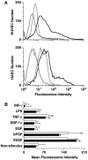
Induction of CXCR4 cell surface expression after stimulation with bFGF and VEGF. A: Flow cytometric analysis of HUVECs and HAECs at 24 hours after stimulation as described in Materials and Methods. Resting endothelial cells: Ab control (broken line) and 12G5 MAb (thin line); VEGF- and bFGF-stimulated endothelial cells: Ab control (gray line) and 12G5 MAb (thick line). B: Mean fluorescence intensity on resting and stimulated HUVECs (filled bars) and HAECs (open bars) at 48 hours after stimulation. The mean and SEM of three experiments is shown. *P < 0.01
Figure 3.
Figure 3.
SDF-1α binds to VEGF- and bFGF-stimulated HUVECs with high affinity. HUVECs were stimulated, and binding of 125I-labeled SDF-1α (1 ng/ml) in the presence of the indicated concentrations of unlabeled SDF-1α was performed as described in Materials and Methods. The bound/total ratio is shown. HUVECs express an average of approximately 16,600 CXCR4 receptors per cell (inset). A representative experiment is shown.
Figure 4.
Figure 4.
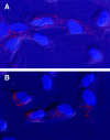
SDF-1α induced CXCR4 redistribution on HUVECs. Cells were stimulated with VEGF (100 ng/ml) for 24 hours. The 12G5 MAb was used to detect CXCR4 as shown in confocal photomicrographs. A: VEGF only. B: VEGF-stimulated cells were washed and exposed to SDF-1α (1 μg/ml for 30 minutes), and redistribution of its receptor was observed.
Figure 5.
Figure 5.
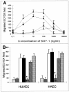
In vitro chemotaxis of HUVECs and HAECs toward SDF-1α is inhibited by CXCR4 antibody. The number of migrated HUVECs and HAECs per 10× field was quantitated as described in Materials and Methods. A: Unstimulated HUVECs (○), VEGF- and bFGF-stimulated HUVECs (•), unstimulated HAECs (▵), VEGF- and bFGF-stimulated HAECs (▴). B: Inhibition of the chemotactic response of HUVECs and HAECs toward SDF-1α (10 ng/ml) by MAb 12G5: Migration was toward medium alone (basal migration; stripes) or in the presence of 12G5 at 10 μg/ml (gray) and migration toward SDF-1α in the absence of antibody (open bars), mouse IgG (10 μg/ml; filled bars ), 12G5 MAb (10 μg/ml; checkerboard bars), 12G5 MAb (1 μg/ml; hatched bars), and 12G5 MAb (0.1 μg/ml; dotted bars); *P < 0.001. The mean and SEM of three experiments are shown.
Figure 6.
Figure 6.
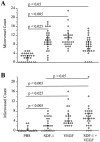
SDF-1α can induce neovascularization in vivo. The number of microvessels is shown per cluster within a section. Ten sections were analyzed per mouse skin section set at day 7 (A) or day 14 (B) after initiation of four daily injections. The mean, SEM and P value for each group of three mice are presented.
Figure 7.
Figure 7.
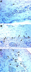
Composite photomicrographs showing neovascularization in SDF-1α-injected mouse skin. Mice received four daily injections of PBS (A), SDF-1 (1 μg; B), or VEGF (1 μg; C) and were biopsied on day 7, and sections were stained for PECAM-1 (CD31), a marker for endothelial cells. Microvessels are indicated by arrows.
Figure 8.
Figure 8.
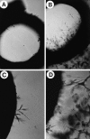
Rat aortic ring assay, showing rat aortic ring capillary sprouting in response to SDF-1α (1 ng/ml). Capillary sprouting occurred from the edge of the ring. A and C: Negative control; B and D: SDF-1α (1 ng/ml). Magnification, ×4 (A and B) and ×40 (C and D). Note that SDF-1α induced formation of long sprouts.
Figure 9.
Figure 9.
SDF-1α-CXCR4 interaction enhanced VEGF release from HUVECs. Cells were cultured as described in Materials and Methods and stimulated for 12 hours with VEGF and bFGF. Thereafter, cells were washed and treated with SDF-1α (1 μg/ml final) for 24 hours, and supernatant samples were collected at 24 hours for ELISA. Open bars, without SDF-1 treatment; hatched bars, with SDF-1α treatment. Concentrations of VEGF produced by different HUVECs are shown. A representative experiment is shown.
Figure 10.
Figure 10.
Hypothetical model of VEGF-, bFGF-, and TNF-α-induced angiogenesis. VEGF or bFGF amplify their angiogenic effects by inducing CXCR4 expression on endothelial cells. TNF-α acts indirectly (**) by inducing the production and release of VEGF and bFGF. CXCR4-positive endothelial cells migrate toward SDF-1α to develop new vessels, and SDF-1-CXCR4 interaction increases VEGF production by endothelial cells, thus amplifying this response.

References

    1. Hanahan D, Folkman J: Patterns and emerging mechanisms of the angiogenic switch during tumorigenesis. Cell 1996, 86:353-364 - PubMed
    1. Ellis LM, Fidler IJ: Angiogenesis and metastasis. Eur J Cancer 1996, 32A:2451-2460 - PubMed
    1. Folkman J: Angiogenesis in cancer, vascular, rheumatoid and other disease. Nature Med 1995, 1:27-31 - PubMed
    1. Auerbach W, Auerbach R: Angiogenesis inhibition: a review. Pharmacol Ther 1994, 63:265-311 - PubMed
    1. Bussolino F, Albini A, Camussi G, Presta M, Viglietto G, Ziche M, Persico G: Role of soluble mediators in angiogenesis. Eur J Cancer 1996, 32A:2401-2412 - PubMed

Publication types

MeSH terms

LinkOut - more resources