. 1999 Jun;73(6):4631-9.
doi: 10.1128/JVI.73.6.4631-4639.1999.

Herpesvirus ateles gene product Tio interacts with nonreceptor protein tyrosine kinases

Affiliations

Herpesvirus ateles gene product Tio interacts with nonreceptor protein tyrosine kinases

J C Albrecht et al. J Virol. 1999 Jun.

Abstract

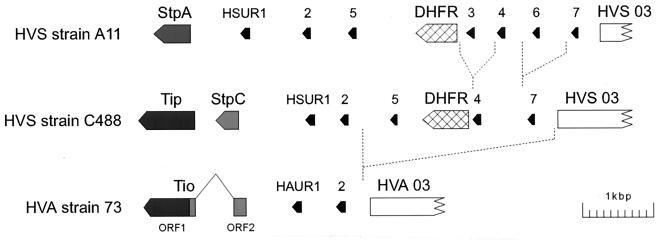
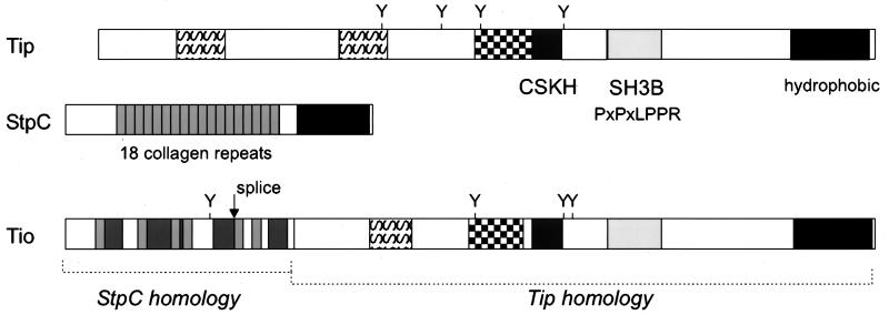
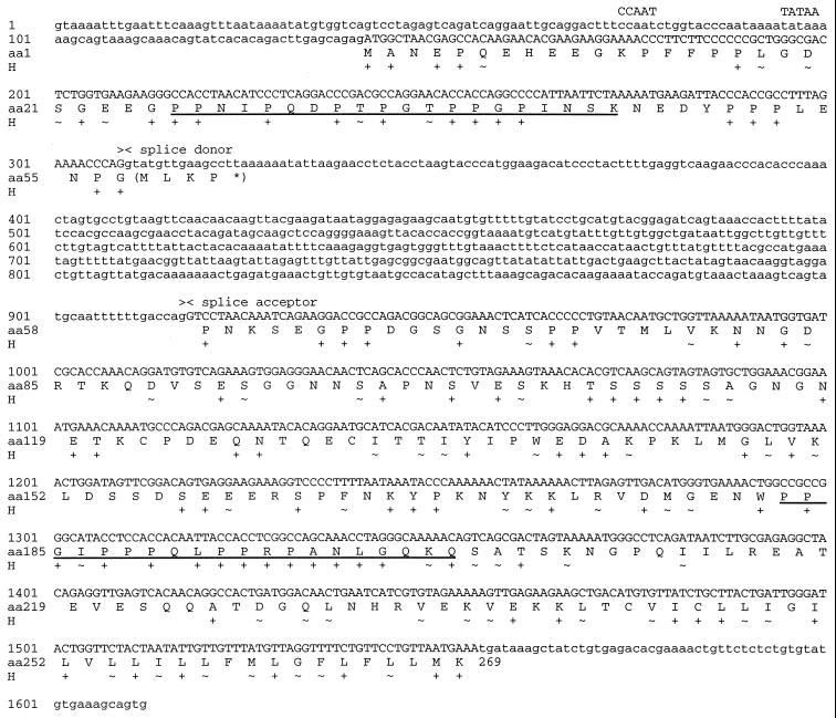
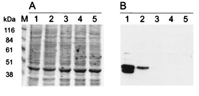
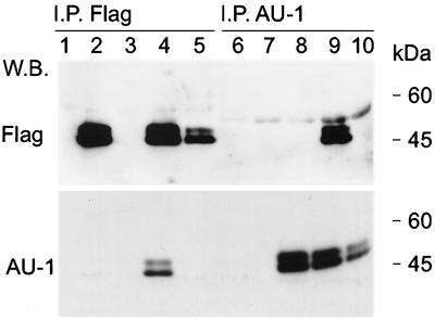
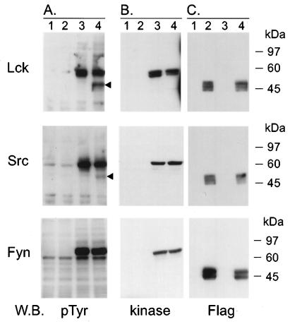
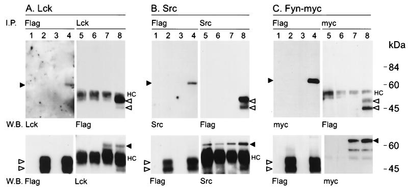
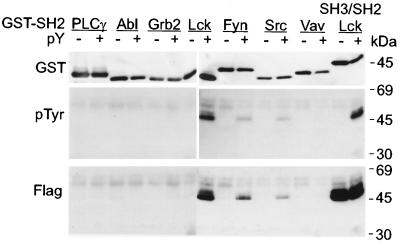
Herpesvirus ateles is a gamma-2-herpesvirus which naturally infects spider monkeys (Ateles spp.) and causes malignant lymphoproliferative disorders in various other New World primates. The genomic sequence of herpesvirus ateles strain 73 revealed a close relationship to herpesvirus saimiri, with a high degree of variability within the left terminus of the coding region. A spliced mRNA transcribed from this region was detected in New World monkey T-cell lines transformed by herpesvirus ateles in vitro or derived from T cells of infected Saguinus oedipus. The encoded viral protein, termed Tio, shows restricted homology to the oncoprotein StpC and to the tyrosine kinase-interacting protein Tip, two gene products responsible for the T-cell-transforming and oncogenic phenotype of herpesvirus saimiri group C strains. Tio was detectable in lysates of the transformed T lymphocytes. Dimer formation was observed after expression of recombinant Tio. After cotransfection, Tio was phosphorylated in vivo by the protein tyrosine kinases Lck and Src and less efficiently by Fyn. Stable complexes of these Src family kinases with the viral protein were detected in lysates of the transfected cells. Binding analyses indicated a direct interaction of Tio with the SH3 domains of Lyn, Hck, Lck, Src, Fyn, and Yes. In addition, tyrosine-phosphorylated Tio bound to the SH2 domains of Lck, Src, or Fyn. Thus, herpesvirus ateles-encoded Tio may contribute to viral T-cell transformation by influencing the function of Src family kinases.


Figures

FIG. 1
FIG. 1
Left-terminal genome variation among strains of HVS and HVA. The variable regions of HVS subgroups A and C encode the proteins StpA, StpC, and Tip, respectively, which are necessary for the transforming phenotype of the virus. Two open reading frames (ORF1 and ORF2) were identified in the corresponding genomic region of HVA strain 73. This region was transcribed into an mRNA of about 1 kb. The transcript was found to be spliced and to code for a single protein, Tio, that has homology to Tip and StpC. The splice occurs within the StpC-homologous portion of the Tio mRNA. Two of the small U RNAs of HVS (HSUR and HAUR) and reading frame 3 (HVS and HVA 03) are conserved. The other HSUR copies and the reading frame for dihydrofolate reductase (DHFR) found in HVS are not present in the genome of HVA.
FIG. 2
FIG. 2
Modular structure of Tio. The Tio molecule can be divided into two sections. The N-terminal third displays 36% amino acid identity to HVS StpC. This homology relies on richness in glycine and proline residues which are also common to collagen repeats. The perfect repetitive collagen-like structure of StpC (small boxes, 30% grey) was not found in Tio. Instead, individual collagen triplets are dispersed between positions 11 and 77 and connected by proline-rich sequences (60% grey boxes). The C-terminal two-thirds of Tio shows 33% identity to Tip. The most prominent feature here is the conservation of the SH3B domain (10% grey) and its functionally associated domain, CSKH (80% grey). The distance between these domains is also conserved. Other Tip-related regions of Tio include a serine-rich motif (S pattern box) and the region around a conserved tyrosine residue (checkerboard pattern box). The hydrophobic C terminus (black box) probably serves as a membrane anchor.
FIG. 3
FIG. 3
Primary structure of the gene for Tio. The presentation is inverted relative to the standard orientation of the HVA strain 73 genome. The nucleotide sequence starts with a region homologous to the StpC promoter and ends with the first nucleotide of the nonrepetitive DNA. The splice donor and acceptor sites of the mRNA are indicated above the nucleotide sequence. The amino acid translation of Tio (amino acids [aa] 1 to 269) is given in single-letter code. The line below the amino acid sequence (H) shows the homology of Tio to StpC and Tip of HVS strain C488. Identical amino acids are marked by plus signs, and similar amino acids are marked by tilde symbols. Similar amino acids were hydrophobic (L, I, V, M, F, Y, and W), basic (R and K), acidic (D and E), polar (N and Q), or small and neutral (G, A, S, and T). The underlined amino acid sequences are those of the synthetic peptides used for fluorescence spectrometry.
FIG. 4
FIG. 4
Expression of Tio in transformed monkey T cells. (A) Coomassie-stained SDS-PAGE gel loaded with 15 μg of total cell lysate of transformed S. oedipus T cells in each lane. Lanes: 1, cell line A661; 2, cell line A1022; 3 to 5, cell lines established with HVS subgroups A, B, and C, respectively. (B) Parallel gel transferred to a PVDF membrane filter and incubated with rabbit anti-Tio serum. Molecular size standards are given on the left.
FIG. 5
FIG. 5
Tio forms homodimers. 293T cells were transfected with expression vectors (lanes 1 and 6), with an expression construct for Flag-Tio (lanes 2 and 7) or AU-Tio (lanes 3 and 8), or with a mixture of both Tio expression plasmids (lanes 4 and 9). In addition, lysates containing either Flag-Tio or AU-Tio were mixed prior to immunoprecipitation (I.P.) (lanes 5 and 10). Precipitations were performed with anti-Flag agarose (lanes 1 to 5) or with anti-AU1 antibody bound to protein A-Sepharose (lanes 6 to 10). Coprecipitated proteins and their controls were detected by Western blotting (W.B.) with anti-Flag (upper panel) or anti-AU1 (lower panel) antibodies. Molecular mass standards are given on the right.
FIG. 6
FIG. 6
Tio is tyrosine phosphorylated by Src family kinases in vivo. 293T cells were transfected with expression vectors (lane 1) or an expression plasmid for Flag-Tio (lane 2), Lck, Src, or myc-tagged Fyn kinase (lane 3) or for Flag-Tio plus the respective kinase (lane 4). Twenty micrograms of total cell lysates was separated by SDS–9% PAGE and analyzed for tyrosine-phosphorylated proteins (A) by Western blotting (W.B.). Expression of the transfected plasmids was controlled by detection of each individual kinase with specific antibodies (B) or, for Flag-Tio, with epitope-specific antibodies (C). Arrowheads to the right of the phosphotyrosine blots indicate a protein detected only after cotransfection of Flag-Tio with Lck or Src. Molecular mass standards are given on the right.
FIG. 7
FIG. 7
Tio coprecipitates with Src family kinases. 293T cells were transfected with expression vectors (lanes 1 and 5) or an expression plasmid for Flag-Tio (lanes 2 and 6), Lck, Src, or myc-tagged Fyn kinase (lanes 3 and 7), or Flag-Tio plus the respective kinase (lanes 4 and 8). Flag-Tio was immunoprecipitated (I.P.) by anti-Flag (lanes 1 to 4) antibody, and Western blotting (W.B.) was performed by using specific antibodies to detect coprecipitated Lck (A), Src (B), or myc-tagged Fyn (C). The reverse experiments were performed by using kinase-specific or anti-myc antibodies for immunoprecipitation (lanes 5 to 8) and anti-Flag antibody for Western blotting. The lower panel shows the results of control Western blotting after reprobing of the membranes with the antibodies used for immunoprecipitation. Open arrowheads, Flag-Tio; black arrowheads, Lck, Src, or Fyn-myc; HC, immunoglobulin heavy chains. Molecular mass standards are given on the right.
FIG. 8
FIG. 8
Tyrosine-phosphorylated Tio binds to GST-SH2 domains of Src family kinases. Flag-tagged Tio fusion proteins were expressed in bacteria with or without an active tyrosine kinase. Both phosphorylated (pY+) and unphosphorylated (pY−) Tio proteins were purified and tested for the ability to bind to GST-SH2 domains derived from the proteins given at the top. Western blotting was performed to control the amount of input GST fusion proteins (GST) and to detect tyrosine-phosphorylated (pTyr), as well as total (Flag), Flag-Tio bound by the GST-SH2 proteins. A GST-SH3/SH2 fusion protein of Lck was used to analyze the binding capacity of the unphosphorylated Tio preparation. Molecular mass standards are given on the right.

References

    1. Albrecht, J. C., and B. Fleckenstein. Unpublished data. GenBank accession no. AF083424.
    1. Albrecht J C, Nicholas J, Biller D, Cameron K R, Biesinger B, Newman C, Wittmann S, Craxton M A, Coleman H, Fleckenstein B. Primary structure of the herpesvirus saimiri genome. J Virol. 1992;66:5047–5058. - PMC - PubMed
    1. Biesinger, B. Unpublished data.
    1. Biesinger B, Muller-Fleckenstein I, Simmer B, Lang G, Wittmann S, Platzer E, Desrosiers R C, Fleckenstein B. Stable growth transformation of human T lymphocytes by herpesvirus samiri. Proc Natl Acad Sci USA. 1992;89:3116–3119. - PMC - PubMed
    1. Biesinger B, Trimble J J, Desrosiers R C, Fleckenstein B. The divergence between two oncogenic Herpesvirus saimiri strains in a genomic region related to the transforming phenotype. Virology. 1990;176:505–514. - PubMed

Publication types

Associated data

LinkOut - more resources