

Quantitative analysis of latent human cytomegalovirus
- PMID: 10233941
- PMCID: PMC112523
- DOI: 10.1128/JVI.73.6.4806-4812.1999
Quantitative analysis of latent human cytomegalovirus
Abstract
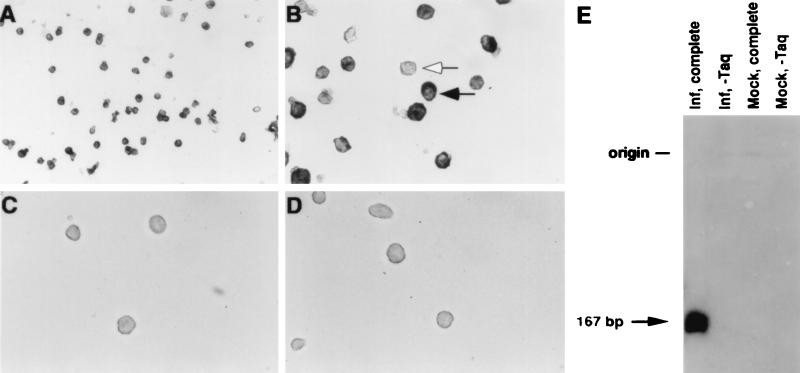
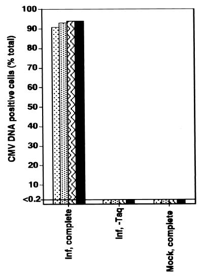
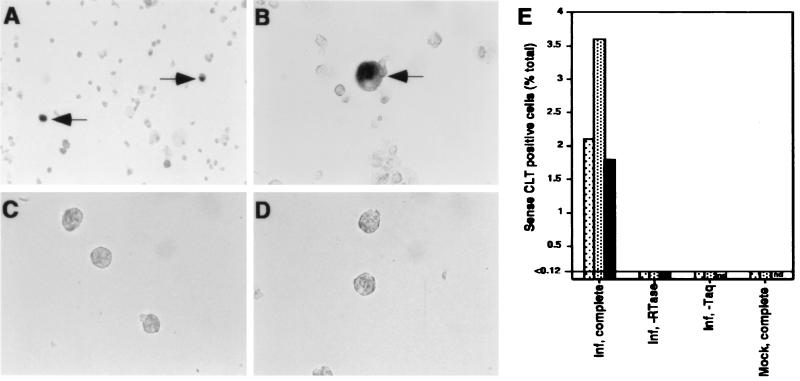
Cytomegalovirus latency depends on an interaction with hematopoietic cells in bone marrow and peripheral blood. The distribution of viral DNA was investigated by PCR-driven in situ hybridization (PCR-ISH), and the number of viral genomes per cell was estimated by quantitative competitive PCR during both experimental and natural latent infection. During experimental latent infection of cultured granulocyte-macrophage progenitors, the viral genome was detected in >90% of cells at a copy number of 1 to 8 viral genomes per cell. During natural infection, viral genomes were detected in 0.004 to 0.01% of mononuclear cells from granulocyte colony-stimulating factor-mobilized peripheral blood or bone marrow from seropositive donors, at a copy number of 2 to 13 genomes per infected cell. When evaluated by reverse transcription-PCR-ISH, only a small proportion of experimentally infected cells (approximately 2%) had detectable latent transcripts. This investigation identifies the small percentage of bone marrow-derived mononuclear cells that become latently infected during natural infection and suggests that latency may proceed in some cells that fail to encode currently identified latent transcripts.
Figures
 , experiment 2;
  ,
experiment 3; ■, experiment 4.
,
experiment 3; ■, experiment 4.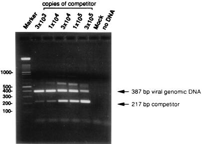
 , experiment 2; ■,
experiment 3.
, experiment 2; ■,
experiment 3.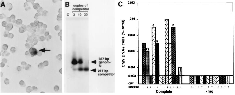
 , mobilized PB 1,
autograft;
, mobilized PB 1,
autograft;  ,
mobilized PB 2, autograft;
,
mobilized PB 2, autograft;
 , mobilized PB 3,
autograft;
, mobilized PB 3,
autograft;  ,
mobilized PB 4, autograft; ■, mobilized PB 5, autograft; ▧,
mobilized PB 6, autograft;
,
mobilized PB 4, autograft; ■, mobilized PB 5, autograft; ▧,
mobilized PB 6, autograft;
 , bone marrow 1,
autograft; ,
mobilized PB 7, allograft; ▨, mobilized PB 8, autograft;
, bone marrow 1,
autograft; ,
mobilized PB 7, allograft; ▨, mobilized PB 8, autograft;
 , mobilized PB 9,
autograft; ▩, mobilized PB 10, autograft;
, mobilized PB 9,
autograft; ▩, mobilized PB 10, autograft;
 , mobilized PB 11,
allograft.
, mobilized PB 11,
allograft.References
-
- Bevan I S, Walker M R, Daw R A. Detection of human cytomegalovirus DNA in peripheral blood leukocytes by the polymerase chain reaction. Transfusion. 1993;33:783–784. . (Letter.) - PubMed
-
- Bolovan-Fritts C A, Mocarski E S, Weideman J A. Peripheral blood CD14+cells from healthy subjects carry a circular conformation of latent cytomegalovirus genome. Blood. 1999;93:394–398. - PubMed
-
- Britt W J, Alford C A. Cytomegalovirus. In: Fields B N, Knipe D M, Howley P M, editors. Fields virology. 3rd ed. Philadelphia, Pa: Lippincott-Raven; 1996. pp. 2493–2523.
Publication types
MeSH terms
Substances
Grants and funding
LinkOut - more resources
Full Text Sources
Other Literature Sources
