. 1999 Jun;73(6):5089-97.
doi: 10.1128/JVI.73.6.5089-5097.1999.

Microglial activation varies in different models of Creutzfeldt-Jakob disease

Affiliations

Microglial activation varies in different models of Creutzfeldt-Jakob disease

C A Baker et al. J Virol. 1999 Jun.

Abstract

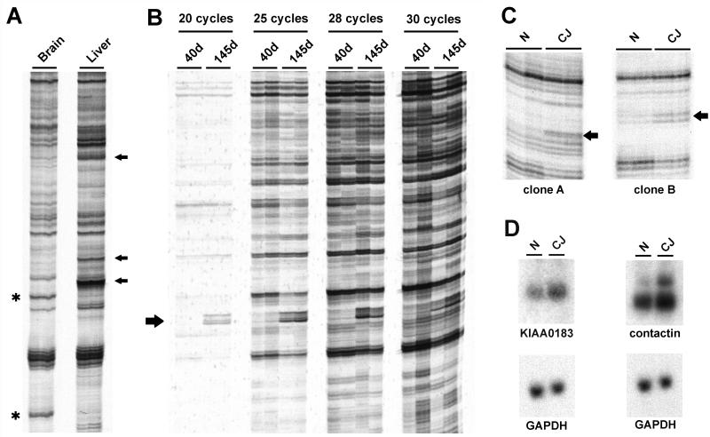
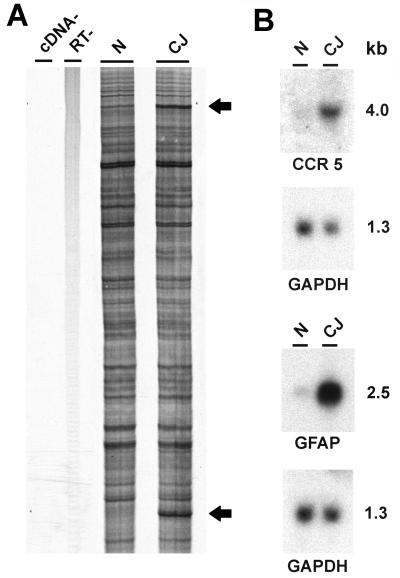
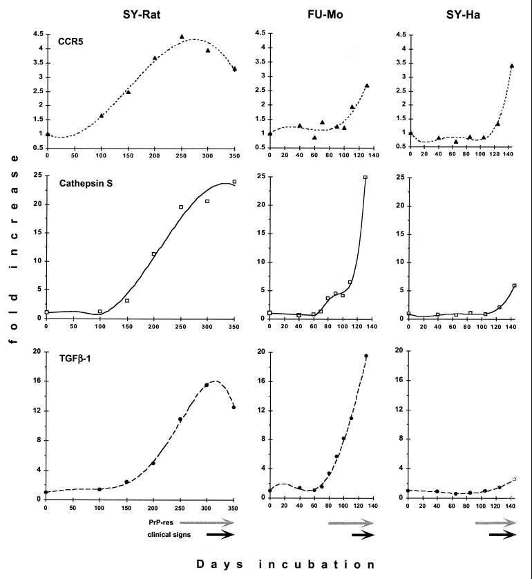
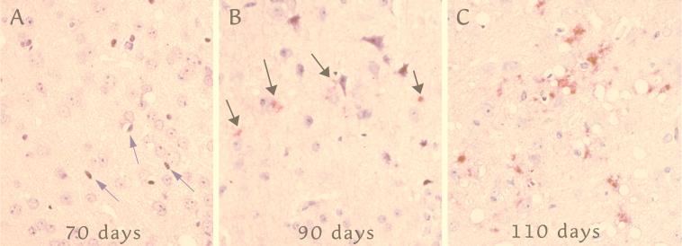
Progressive changes in host mRNA expression can illuminate crucial pathogenetic pathways in infectious disease. We examined general and specific approaches to mRNA expression in three rodent models of Creutzfeldt-Jakob disease (CJD). Each of these models displays distinctive neuropathology. Although mRNAs for the chemokine receptor CCR5, the lysosomal protease cathepsin S, and the pleiotropic cytokine transforming growth factor beta1 (TGF-beta1) were progressively upregulated in rodent CJD, the temporal patterns and peak magnitudes of each of these transcripts varied substantially among models. Cathepsin S and TGF-beta1 were elevated more than 15-fold in mice and rats infected with two different CJD strains, but not in CJD-infected hamsters. In rats, an early activation of microglial transcripts preceded obvious deposits of prion protein (PrP) amyloid. However, in each of the three CJD models, the upregulation of CCR5, cathepsin S, and TGF-beta1 was variable with respect to the onset of PrP pathology. These results show glial cell involvement varies as a consequence of the agent strain and species infected. Although neurons are generally assumed to be the primary sites for agent replication and abnormal PrP formation, microglia may be targeted by some agent strains. In such instances, microglia can both process PrP to become amyloid and can enhance neuronal destruction. Because microglia can participate in agent clearance, they may also act as chronic reservoirs of infectivity. Finally, the results here strongly suggest that TGF-beta1 can be an essential signal for amyloid deposition.


Figures

FIG. 1
FIG. 1
DD of control and CJD samples. (A) cDNA samples in duplicate from hamster brain and liver showing typical analysis with a standard arbitrary primer pair. Several brain-enriched and liver-enriched DD products are indicated by asterisks and arrows, respectively. (B) DD using primers targeted to the hamster GFAP cDNA sequence. RNA samples in duplicate from CJD-infected hamsters 40 and 145 days after inoculation were amplified by RT-PCR for 20, 25, 28, or 30 PCR cycles. Each PCR was performed in duplicate to verify reproducibility of the PCR products. The 192-bp GFAP product is indicated by an arrow. (C) DD of normal (N) and terminal CJD (CJ) of the SY-Ha model verified in duplicate. Differentially expressed products are indicated by arrows. (D) Northern blots of total RNA from normal and terminal SY-Ha CJD confirm differential expression compared to that of the steady-state GAPDH control.
FIG. 2
FIG. 2
Directed DD and confirmation of CCR5 upregulation in SY-Ha CJD. (A) Degenerate primers directed to chemokine receptor motifs were used to amplify normal (N) and terminal CJD (CJ) hamster cDNA. The upper and lower bands (arrows) correspond to CCR5 and GFAP, respectively. The GFAP band had been amplified from the 3′-untranslated region of the transcript by the degenerate primers. No bands are seen in the two negative control experiments performed in parallel, one without cDNA (cDNA− lane) and the second without reverse transcriptase (RT− lane). (B) Northern blots of total hamster brain RNA confirm upregulation of sequences identified by DD. Approximate molecular weights (kilobases) are indicated.
FIG. 3
FIG. 3
Time course of gene expression in the SY-Rat CJD model. Northern blots of total rat brain RNA isolated at the indicated times (days [d]) after inoculation were sequentially hybridized with the CCR5, cathepsin S, TGF-β1, and GAPDH probes. Hamster and mouse Northern blots were analyzed in the same manner (data not shown). GAPDH expression was used as a control to normalize for sample loading.
FIG. 4
FIG. 4
Quantitative analysis of Northern blot transcripts showing marked differences between models. Each time point represents a pooled RNA sample from two animal brains. CCR5 (upper row, solid triangles), cathepsin S (middle row, open squares), and TGF-β1 (lower row, solid circles) are shown in the SY-Rat, FU-Mo, and SY-Ha models of CJD. Values are normalized to levels of GAPDH mRNA and expressed in terms of the fold increase over the corresponding normal brain sample (indicated here as day 0). The curve fits shown for mRNA increases all have r2 values greater than 0.977, except for CCR5 expression in the FU-Mo model (r2 = 0.896). Note the different scales for the expression levels of the three transcripts. PrP-res levels and clinical signs were assessed with the same animals used for these RNA studies. The onset of major PrP-res accumulation is indicated by gray arrows, and the durations of clinical symptoms are indicated by black arrows as previously documented in the SY-Rat and SY-Ha models (35, 36), with PrP-res increasing ∼20-fold after 200 days in SY-Rat infection and >30-fold after 87 days in SY-Ha infection. Figure 5 documents the major deposition of PrP amyloid in the FU-Mo model after 80 days.
FIG. 5
FIG. 5
Maximal levels of PrP-res in representative cortical sections taken every 10 days in the FU-Mo model (see text). (A) At 70 days, a few elongated nuclei typical of microglia (arrows) are seen with very rare vacuoles and no detectable PrP-res. (B) A rare focus at 90 days showing few small, granular, abnormal PrP deposits (red; arrows). (C) By 110 days, vacuoles and abundant larger deposits of abnormal PrP are seen. The sections were stained in parallel as described in reference and were counterstained with hematoxylin.

References

    1. Aguzzi A, Weissmann C. Prion research: the next frontiers. Nature. 1997;389:795–798. - PubMed
    1. Akowitz A, Sklaviadis T, Manuelidis E E, Manuelidis L. Nuclease-resistant polyadenylated RNAs of significant size are detected by PCR in highly purified Creutzfeldt-Jakob disease preparations. Microb Pathog. 1990;9:33–45. - PubMed
    1. Akowitz A, Sklaviadis T, Manuelidis L. Endogenous viral complexes with long RNA cosediment with the agent of Creutzfeldt-Jakob disease. Nucleic Acids Res. 1994;22:1101–1107. - PMC - PubMed
    1. Berglund E O, Ranscht B. Molecular cloning and in situ localization of the human contactin gene (CNTN1) on chromosome 12q11-q12. Genomics. 1994;21:571–582. - PubMed
    1. Bruce M E, McConnell I, Fraser H, Dickinson A G. The disease characteristics of different strains of scrapie in Sinc congenic mouse lines: implications for the nature of the agent and host control of pathogenesis. J Gen Virol. 1991;72:595–603. - PubMed

Publication types