. 1999 May 15;19(10):3681-90.
doi: 10.1523/JNEUROSCI.19-10-03681.1999.

Encephalopsin: a novel mammalian extraretinal opsin discretely localized in the brain

Affiliations

Encephalopsin: a novel mammalian extraretinal opsin discretely localized in the brain

S Blackshaw et al. J Neurosci. .

Abstract

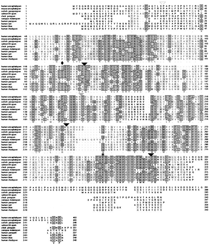
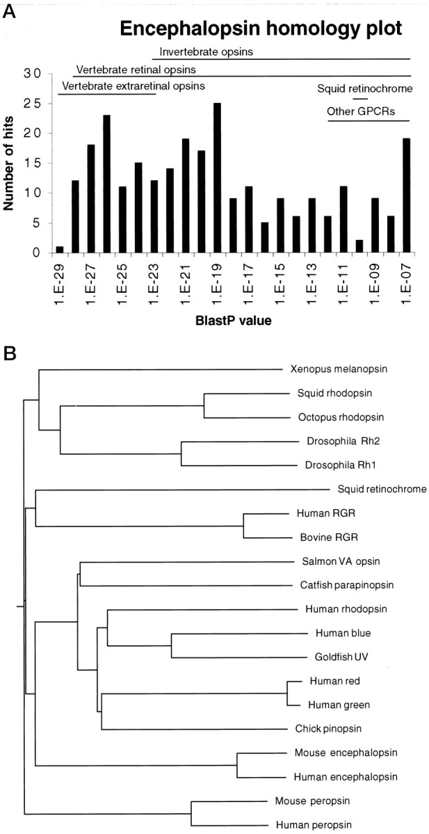
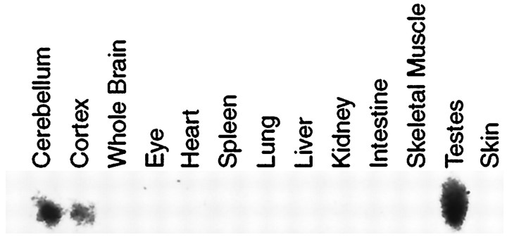
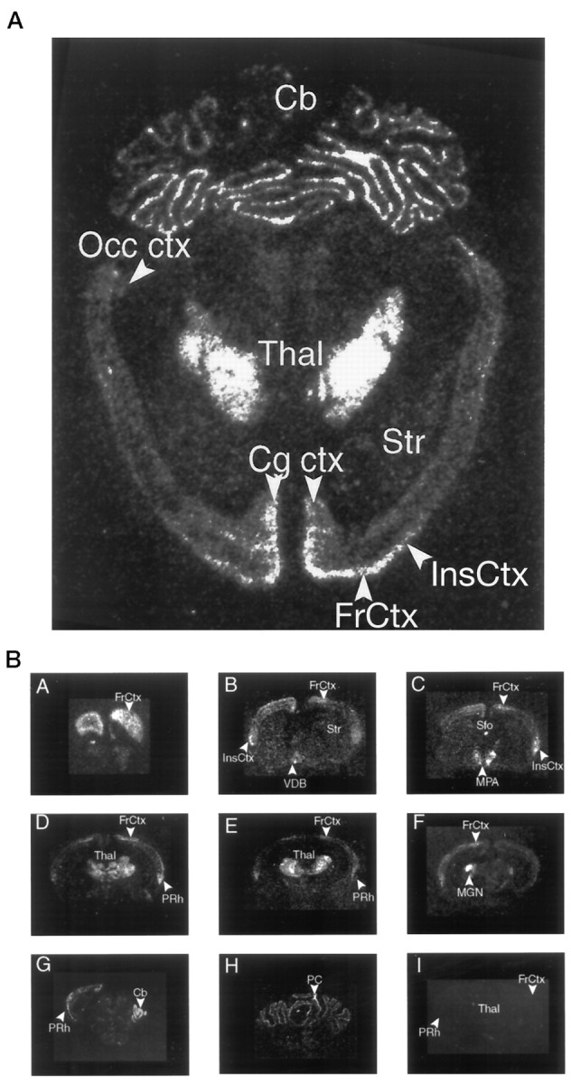
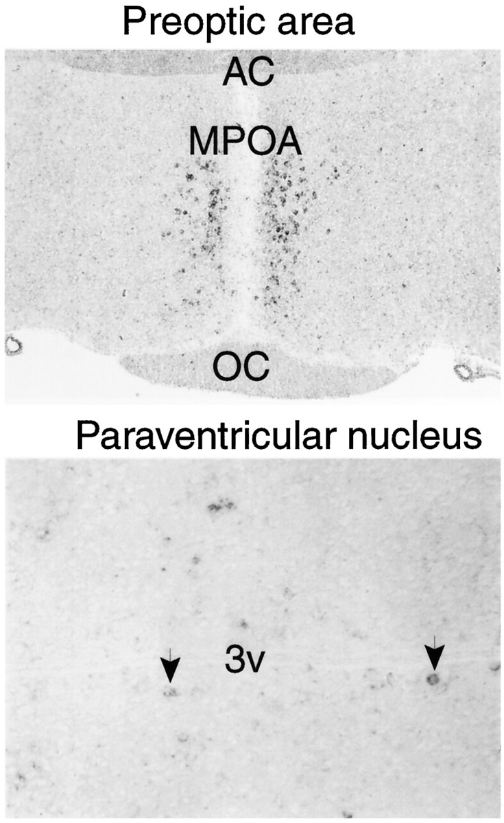
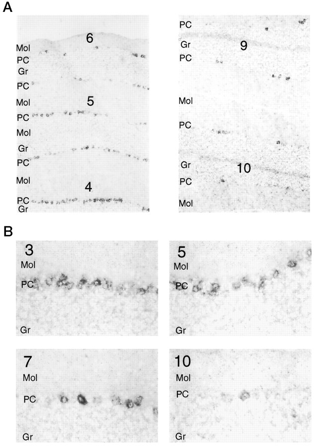
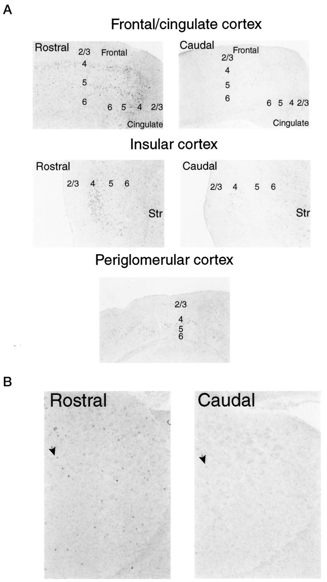
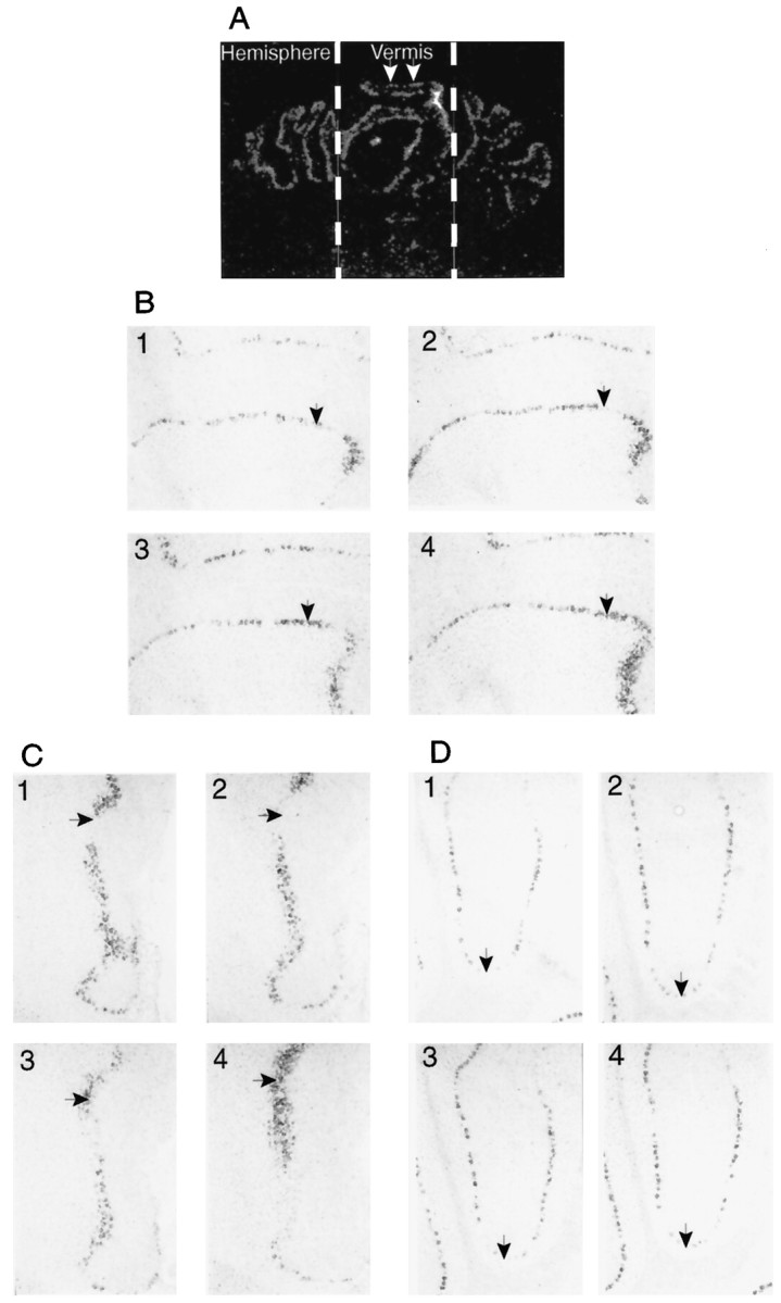
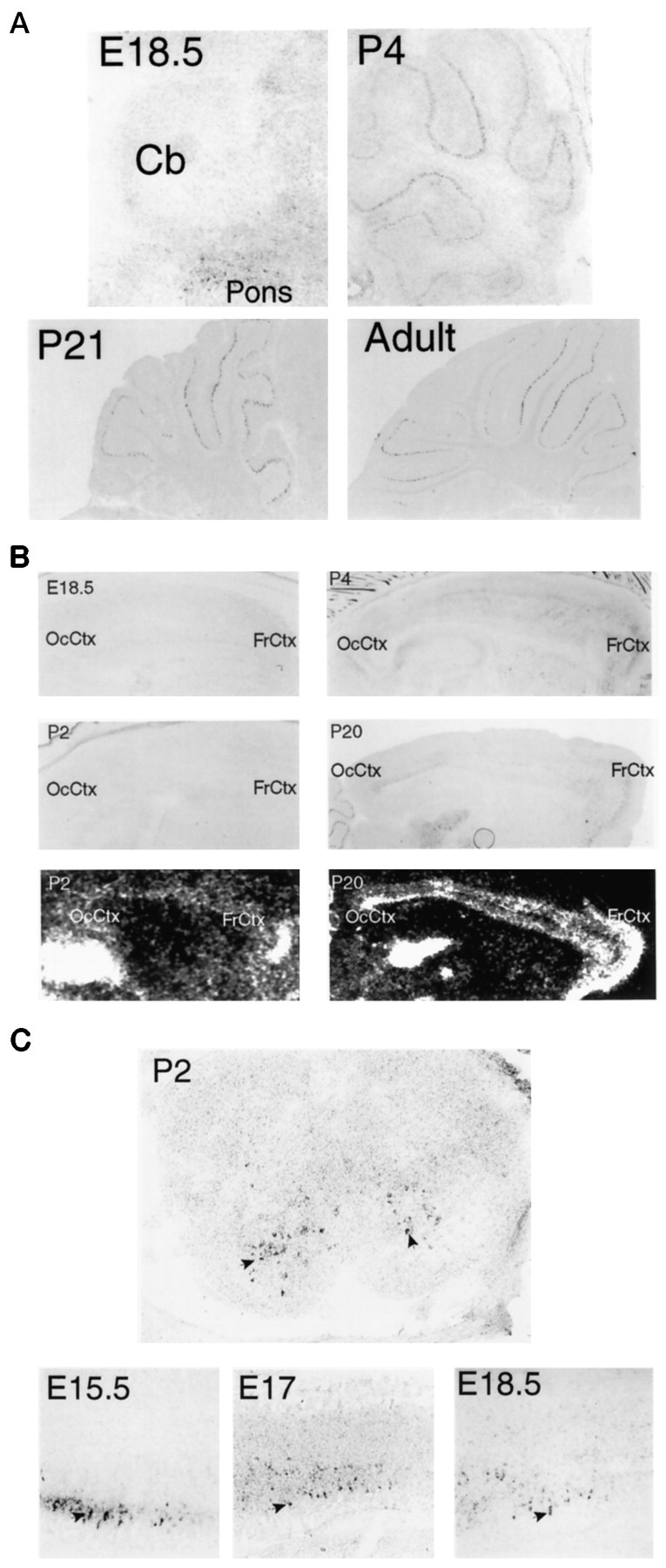
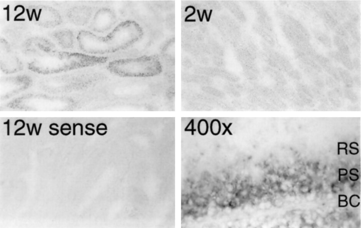
We have identified a mammalian opsin, encephalopsin, that shows strong and specific expression in the brain. Encephalopsin defines a new family of opsins and shows highest homology to vertebrate retinal and pineal opsins. Encephalopsin is highly expressed in the preoptic area and paraventricular nucleus of the hypothalamus, both regions implicated in encephalic photoreception in nonmammalian vertebrates. In addition, encephalopsin shows highly patterned expression in other regions of the brain, being enriched in selected regions of the cerebral cortex, cerebellar Purkinje cells, a subset of striatal neurons, selected thalamic nuclei, and a subset of interneurons in the ventral horn of the spinal cord. Rostrocaudal gradients of encephalopsin expression are present in the cortex, cerebellum, and striatum. Radial stripes of encephalopsin expression are seen in the cerebellum. In the cortex and cerebellum, encephalopsin expression is considerably higher and more highly patterned in the adult than in the neonate. Encephalopsin is the first putative extraocular opsin identified in mammals and may play a role in encephalic photoreception.


Figures

Fig. 1.
Fig. 1.
Primary sequence of human and mouse encephalopsin. Sequences are aligned via CLUSTALW with members of each of the main classes of vertebrate retinal and extraretinal opsins. Residues conserved in the majority of sequences are in darkly shaded boxes, whereas similar residues are in lightly shaded boxes. The position of the lysine in the seventh transmembrane domain that forms a Schiff base with retinal is indicated by anasterisk, whereas the position of the counterion in the third transmembrane domain is indicated by a diamond. Positions of the introns present in encephalopsin and the rod, cone, and pineal opsins are indicated with a black arrowhead. The gray arrowhead indicates the intron that is present in rod and cone opsins, along with pinopsin, but is absent in encephalopsin and parapinopsin. The white arrowheadindicates the position of the N-terminal intron found in the red- and green-sensitive opsins.
Fig. 2.
Fig. 2.
A, P value distribution of matches to mouse encephalopsin matches determined by a BLASTP search using BLAST 2.0 against the December 1998 GenBank release. Redundant sequences were not eliminated for this analysis. Other GPCRs represent nonopsin G-protein-coupled receptors. B, Dendrogram of encephalopsin phylogeny. Dendrograms are determined by CLUSTALW analysis. The gap penalty is set at 2, and the gap extension penalty is set at 0.01. Encephalopsin groups with vertebrate retinal and pineal opsins.
Fig. 3.
Fig. 3.
Northern blot analysis of mouse encephalopsin expression. A single band of 1.9 kb is detected.
Fig. 4.
Fig. 4.
A, Expression of encephalopsin in a horizontal section of adult mouse brain. Radioactive in situ hybridization is used. B, Expression of encephalopsin in a rostrocaudal series of coronal sections of adult mouse brain. Radioactive in situ hybridization is used.Cb, Cerebellum; Cg ctx, cingulate cortex;FrCtx, frontal cortex; InsCtx, insular cortex; MPA, medial preoptic area; Occ ctx, occipital cortex; PC, Purkinje cell of cerebellum; PRh, perirhinal cortex; Sfo, subfornical organ; Str, striatum; Thal, thalamus; VDB, ventral diagonal band.
Fig. 5.
Fig. 5.
Digoxygenin in situ hybridization of encephalopsin expression in diencephalic structures.Top, The preoptic nucleus is taken at 50×.Bottom, The paraventricular nucleus is at 200×.Arrows indicate selected encephalopsin-positive cells in the paraventricular nucleus of the hypothalamus. AC, Anterior commissure; MPOA, medial preoptic area;OC, optic chiasm; 3v, third ventricle.
Fig. 6.
Fig. 6.
A, Encephalopsin shows a rostrocaudal gradient of expression in the cerebellum. Digoxygeninin situ hybridization is used. Numbersindicate the lobe of the vermis and are numbered in rostrocaudal order. Pictures are taken at 50×. B, Both the number of encephalopsin-expressing cells and the intensity of encephalopsin expression show a rostrocaudal gradient in the cerebellum. Pictures are taken at 400×. Gr, Granule cell layer;Mol, molecular layer; PC, Purkinje cell layer.
Fig. 7.
Fig. 7.
A, Encephalopsin expression shows a rostrocaudal gradient within the cerebral cortex. Cortical layers are indicated by numbers. Digoxygenin in situhybridization is used. All pictures are taken at 50×.B, Encephalopsin expression shows a rostrocaudal expression gradient in the striatum. Arrows indicate selected encephalopsin-positive cells. Pictures are taken at 50×.Str, Striatum.
Fig. 8.
Fig. 8.
A, Encephalopsin is expressed in a striped pattern in the cerebellum. Radioactive in situhybridization is used on coronal sections. The vermis and hemispheres are indicated. White arrows indicate two consecutive stripes. B, Horizontal stripes of encephalopsin expression in the vermis are shown. Digoxygenin in situhybridization is used here. Consecutive sections are shown and numbered accordingly. All pictures are taken at 50×. C, Vertical stripes of encephalopsin expression are seen in the vermis. The pictures shown are from the same adjacent sections used inB. All pictures are taken at 50×. D, Stripes of encephalopsin expression in the cerebellar hemispheres are shown. Pictures are taken from the same series of consecutive sections used in B and C. All pictures are taken at 50×.
Fig. 9.
Fig. 9.
A, Developmental expression of encephalopsin in the cerebellum is shown. The adult pattern of encephalopsin expression is not seen until several weeks after birth. Sagittal sections are shown. E18.5 is taken at 100×, P4 is taken at 50×, and P21 and adult are taken at 25×. B, Developmental expression of encephalopsin expression in the cerebral cortex is shown. Encephalopsin expression is absent prenatally, relatively evenly distributed throughout the cortex in the early postnatal mouse, and showing rostrocaudal gradients and regional organization by P20. Digoxy genin in situ hybridization is shown for E18.5, P2, P4, and P20, whereas radioactive in situhybridization (bottom) is shown for P2 and P20. Digoxygenin in situ hybridization pictures are taken at 50× for E18.5, 25× for P2 and P4, and 12.5× for P20.C, Encephalopsin expression is prominent in developing spinal cord. Sagittal sections of embryonic time points are taken of the cervical cord at 100× for E15.5 and 50× for E17 and E18.5. The coronal section at P2 is taken of cervical spinal cord at 50×.Arrows indicate selected encephalopsin-positive interneurons of the ventral horn. The color reaction in the P2 section is allowed to proceed twice as long as that in the embryonic time points, so the observed abundance levels of encephalopsin are not directly comparable. Cb, Cerebellum;FrCtx, frontal cortex; OcCtx, occipital cortex.
Fig. 10.
Fig. 10.
Strong encephalopsin expression is observed in meiotic spermatids. Encephalopsin expression is seen in a subset of seminiferous tubules of the 12-week-old (12w) but not the 2 week-old (2w) mouse, indicating expression in sperm. High-power pictures indicate that encephalopsin expression is restricted to pachytene spermatids. All pictures are at 100×, except for the high-power shot that is at 400×. BC, Basal cells; PS, pachytene spermatids; RS, round spermatids.

References

    1. Balsalobre A, Damiola F, Schibler U. A serum shock induces circadian gene expression in mammalian tissue culture cells. Cell. 1998;93:929–937. - PubMed
    1. Blackshaw S, Snyder SH. Developmental expression pattern of phototransduction components in mammalian pineal implies a light-sensing function. J Neurosci. 1997a;17:8074–8082. - PMC - PubMed
    1. Blackshaw S, Snyder SH. Parapinopsin, a novel catfish opsin localized to the parapineal organ, defines a new gene family. J Neurosci. 1997b;17:8083–8092. - PMC - PubMed
    1. Campbell SS, Murphy PJ. Extraocular circadian phototransduction in humans. Science. 1998;279:396–399. - PubMed
    1. Cepko CL. The patterning and onset of opsin expression in vertebrate retinae. Curr Opin Neurobiol. 1996;6:542–546. - PubMed

Publication types

MeSH terms

Associated data