. 1999 May 15;19(10):3761-72.
doi: 10.1523/JNEUROSCI.19-10-03761.1999.

The ankle-link antigen: an epitope sensitive to calcium chelation associated with the hair-cell surface and the calycal processes of photoreceptors

Affiliations

The ankle-link antigen: an epitope sensitive to calcium chelation associated with the hair-cell surface and the calycal processes of photoreceptors

R Goodyear et al. J Neurosci. .

Erratum in

  • J Neurosci 1999 Aug 15;19(16):7238-9

Abstract

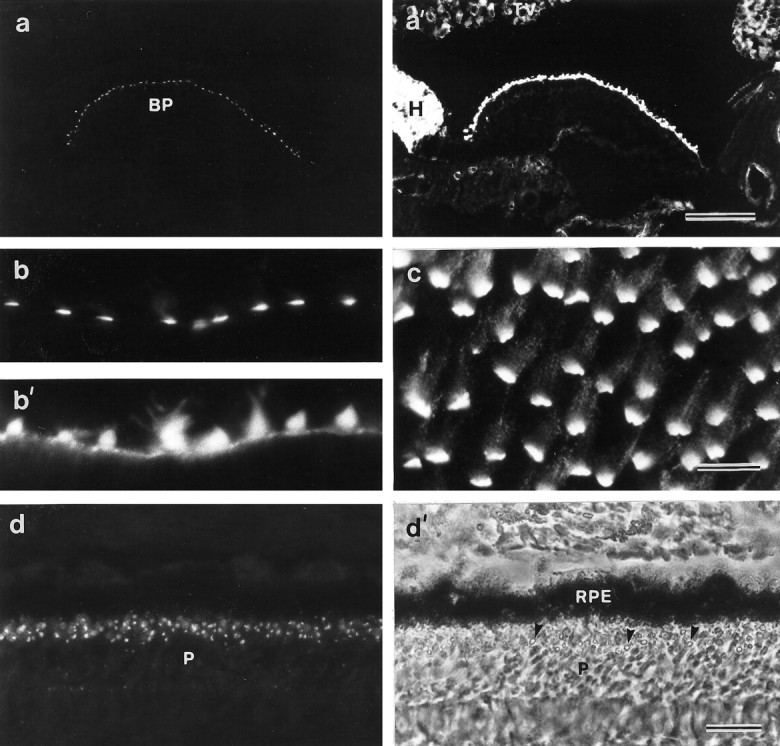
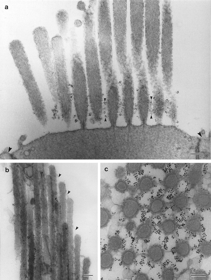
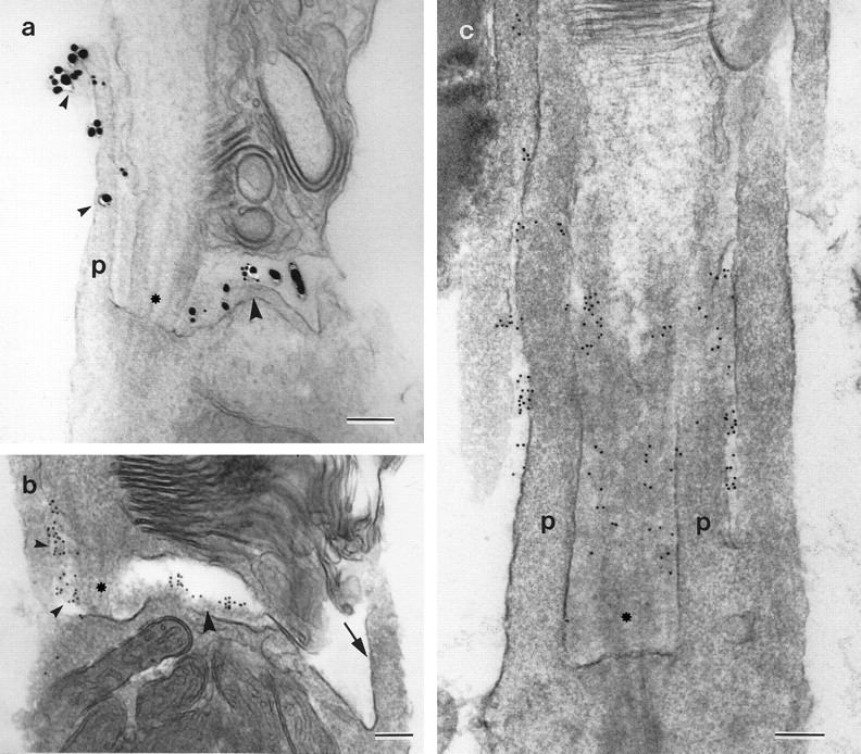
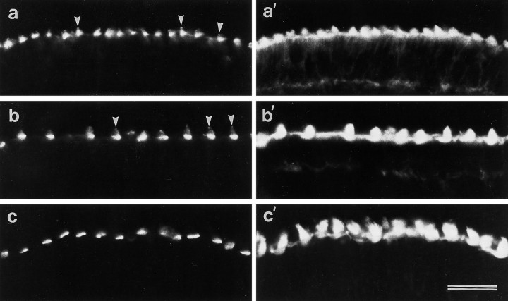
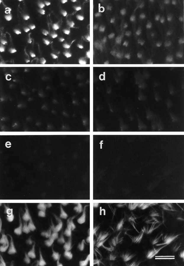
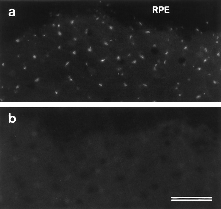
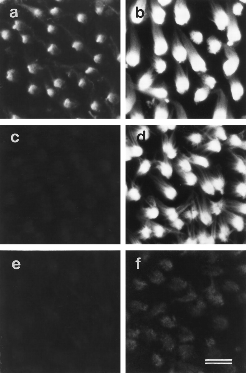
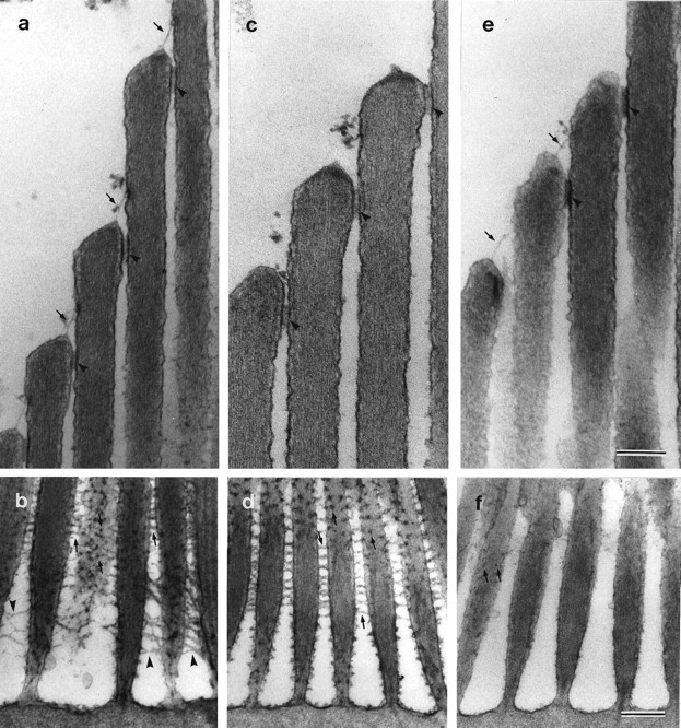
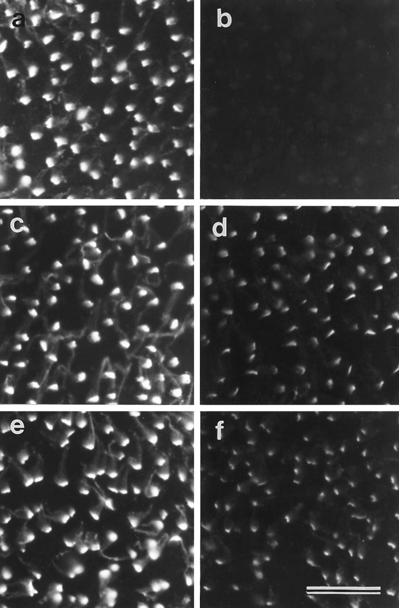
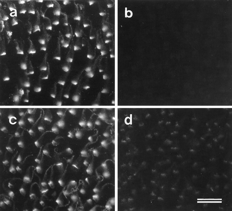
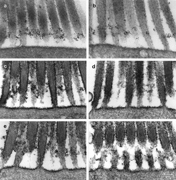
A monoclonal antibody, mAb E40, that specifically recognizes hair cells and photoreceptors was derived from a mouse immunized with a membrane fraction prepared from the sensory maculae of the chick inner ear. In the mature chick inner ear, punctate labeling is observed along each stereocilium, but staining is mostly concentrated around the basal end of the sensory hair bundles, where it is closely associated with surface specializations known as ankle links. The epitope recognized by mAb E40 is therefore referred to as the ankle-link antigen (ALA). During early embryogenesis, the ALA is initially distributed evenly over the surface of the hair bundle. As development proceeds, it becomes more restricted to the base of the hair bundle, although a spot of the ALA remains associated with the bundle tip until just before hatching. In the eye, mAb E40 stains the calycal processes of photoreceptors. When maculae and retinae are treated with the calcium chelator BAPTA at room temperature, the ALA disappears. BAPTA-induced loss of the ALA from the hair-bundle surface is substantially reduced by lowering the temperature to 2 degrees C. The ALA and ankle links reappear on the hair-bundle surface when cells are cultured for 20 hr after BAPTA treatment. BAPTA sensitivity and recovery after BAPTA-induced loss are properties similar to those described for the tip link, a surface structure thought to gate the mechanotransducer channel. However, unlike the tip link, the ALA and ankle links are sensitive to subtilisin treatment. The results define a new component of the hair-bundle surface, with properties both common to and distinct from those of the tip link.


Figures

Fig. 1.
Fig. 1.
Localization of mAb E40 binding sites in the inner ear and the retina. a, a′, Section of the cochlear duct double-labeled with mAb E40 (a) and phalloidin (a′). Within the cochlear duct, mAb E40 is specific for the apical surface of the hair cells in the basilar papilla. Scale bar, 100 μm. b, b′, High-magnification image of a cryosection from the utricular macula double-labeled with mAb E40 (b) and phalloidin (b′). In sectioned material, mAb E40 staining is predominantly restricted to the base of the hair bundle.c, A whole-mount preparation of the utricular macula labeled with mAb E40. Although the majority of the labeling is restricted to the base of each hair bundle, punctate staining can be seen all the way up the stereocilia. Scale bar: b,b′, c, 10 μm. d,d′, Section of the retina immunolabeled with mAb E40 (d) and the corresponding phase-contrast image (d′). A spot of label is associated with each photoreceptor at a level close to that of the oil droplets in the cones. Arrowheads in d′ indicate oil droplets. Scale bar, 20 μm. BP, Basilar papilla;H, homogene cells; P, photoreceptors;RPE, retinal pigment epithelium; TV, tegmentum vasculosum.
Fig. 2.
Fig. 2.
Ultrastructural localization of mAb E40 binding sites on hair cells. a, Striolar hair bundle from the utricular macula labeled with mAb E40 and 10 nm gold-conjugated anti-mouse IgG. Labeling is concentrated around the basal region of the hair bundle, where it is closely associated with the ankle links (small arrowheads). Large arrowheadsindicate adjacent supporting cell surfaces. b, Extrastriolar hair bundle from the utricular macula. Labeling is seen in discrete spots along stereocilia, but the tip links (arrowheads) are unlabeled (200-nm-thick section).c, Section cut parallel to the apical surface of a striolar hair cell from the utricular macula at the level of the ankle links. The ankle links are heavily labeled. Samples in band c were labeled with mAb E40, rabbit anti-mouse IgG1, and 10 nm gold-conjugated goat anti-rabbit IgG. Scale bars: a, 300 nm; b,c, 200 nm.
Fig. 3.
Fig. 3.
Ultrastructural localization of mAb E40 binding sites on rod photoreceptors. The connecting cilium is indicated byasterisks, and p indicates the calycal processes. a, A receptor labeled with mAb E40 and 1 nm gold-conjugated goat anti-mouse IgG, followed by silver intensification. Staining is seen along the calycal process (small arrowheads) projecting from the inner segment adjacent to the connecting cilium and in the region just below the disk stack (large arrowhead). b,c, Photoreceptors labeled with mAb E40, rabbit anti-mouse IgG1, and 10 nm gold-conjugated goat anti-rabbit IgG. In b, gold particles are visible between the calycal process and the connecting cilium (small arrowheads) and in the gap between the inner and outer segments (large arrowhead). Note how the calycal process on the side opposite to the connecting cilium (arrow) is unlabeled. In c, the section grazes the connecting cilium, revealing gold particles in the space lying between the connecting cilium and the calycal processes. Label is also seen between adjacent calycal processes. Scale bars, 200 nm.
Fig. 4.
Fig. 4.
Distribution of mAb E40 binding sites and F-actin during development of the basilar papilla. Cryosections from papillae at the E12 (a, a′), E16 (b, b′), and 2 d after hatch (c, c′) stages of development double-labeled with mAb E40 (a,b, c) and phalloidin (a′,b′, c′). a,a′, At E12, mAb E40 staining (a) is distributed evenly over most of the bundle, although the tips of many hair bundles are decorated by a distinct spot of mAb E40 staining (arrowheads). b, b′, At E16, mAb E40 staining (b) is more concentrated at the base of the bundle, although spots of staining can be observed at the tips of many bundles (arrowheads). c,c′, At 2 d after hatching, mAb E40 staining (c) is primarily restricted to the base of the bundles, and the spots of staining seen at the tip of the bundles at earlier stages of development are no longer visible. Scale bar, 20 μm.
Fig. 5.
Fig. 5.
Effects of BAPTA treatment on mAb E40 labeling in extrastriolar regions of the utricular macula. a, Hair cells in a control macula incubated for 1 hr in HBHBSS before fixation and labeling with mAb E40. Strong, basally concentrated mAb E40 labeling is seen on the hair bundles. b–f, Hair cells in maculae treated with 5 mm BAPTA for 10 sec (b), 1 min (c), 10 min (d), 20 min (e), and 1 hr (f) before fixation and labeling with mAb E40. Staining is considerably reduced after 10 sec (b) and completely eliminated after 1 hr of BAPTA treatment (f). g, h, Phalloidin double labels of the images in a andf, respectively. Note in h how the stereocilia in the hair bundles that have been treated with BAPTA for 1 hr are splayed. Scale bar, 10 μm.
Fig. 6.
Fig. 6.
Effects of BAPTA treatment on mAb E40 labeling in retina whole mounts. a, Control retina incubated in HBHBSS for 20 min before fixation and labeling with mAb E40. Punctate staining of the photoreceptors is observed in areas from which the retinal pigment epithelium (RPE) has been removed before treatment. b, Retina incubated in 5 mm BAPTA for 20 min before fixation and labeling with mAb E40. Note the loss of labeling. The dark area across the top of each micrograph is a region covered by the retinal pigment epithelium. Scale bar, 20 μm.
Fig. 7.
Fig. 7.
Effects of BAPTA and subtilisin on mAb E40 (a, c, e) and anti-HCA labeling (b, d, f) in extrastriolar regions of utricular maculae. a,b, Control samples incubated in HBHBSS for 60 min.c, d, Samples incubated in 5 mm BAPTA for 60 min. e, f, Samples incubated in 50 μg/ml subtilisin for 20 min. In control maculae (a, b), both mAb E40 (a) and monoclonal anti-HCA antibody (b) label hair bundles strongly. After 1 hr of BAPTA treatment, mAb E40 antigen no longer stains the cell surface (c), whereas the distribution of the hair-cell antigen is unchanged (d), although the hair bundles are splayed. After 20 min exposure to subtilisin, the mAb E40 staining can no longer detected (e), and only traces of the HCA remain (f). Scale bar, 10 μm.
Fig. 8.
Fig. 8.
Effects of BAPTA and subtilisin on hair-bundle links and connectors. Ultrastructural appearance of the upper regions of tannic acid-stained striolar hair bundles (a,c, e) and the basal regions of ruthenium red-stained extrastriolar hair bundles (b,d, f) in utricular maculae incubated with HBHBSS (a, b), BAPTA (c, d), and subtilisin (e,f). In a, c, ande, arrows indicate tip links, andarrowheads indicate horizontal top connectors. Inb, d, and f,arrows indicate shaft connectors, andarrowheads indicate ankle links. a,b, In controls incubated in HBHBSS for 20 min, tip links, horizontal top connectors, shaft connectors, and ankle links can all be seen. c, d, After treatment with 5 mm BAPTA for 20 min, tip links and ankle links can no longer be observed, but horizontal top links and shaft links are still visible. e, f, After treatment with 50 μg/ml subtilisin for 20 min, tip links and horizontal top links are still present, only a few remnants of the shaft links remain, and ankle links are absent. Scale bars: a–f, 300 nm.
Fig. 9.
Fig. 9.
Temperature dependence of the effects of BAPTA treatment on mAb E40 labeling in extrastriolar regions of the utricular macula. a, b, Maculae stained with mAb E40 after a 20 min incubation in HBHBSS (a) or 5 mm BAPTA (b) at room temperature.c, d, Maculae stained with mAb E40 after a 20 min incubation in HBHBSS (c) or 5 mm BAPTA (d) at 2°C. Although staining is reduced in the tissue treated with BAPTA at 2°C compared with the control tissue, labeling is much stronger than that seen after BAPTA treatment at room temperature (b).e, f, Maculae stained with mAb E40 after a 4 hr incubation in HBHBSS (e) or 5 mm BAPTA (f) at 2°C. The degree of labeling observed after BAPTA treatment for 4 hr at 2°C (f) is similar to that seen after 20 min at 2°C (d). Scale bar, 20 μm.
Fig. 10.
Fig. 10.
Recovery of mAb E40 labeling after BAPTA-induced loss. a, Control macula stained with mAb E40 after 20 min incubation in HBHBSS. b, BAPTA-treated (5 mm, 20 min) macula stained with mAb E40. Note that only very weak labeling can be detected. c, Control, HBHBSS-treated macula after 20 hr in vitro stained with mAb E40. d, BAPTA-treated (5 mm, 20 min) macula stained with mAb E40 after 20 hr in vitro. mAb E40 staining has reappeared but not to the same level as in the control (b). Scale bar, 10 μm.
Fig. 11.
Fig. 11.
Ankle links and mAb E40 binding sites on extrastriolar hair cells from the utricular macula. a,b, Effects of BAPTA at 2°C. Hair bundles from maculae incubated in HBHBSS (a) or 5 mm BAPTA (b) at 2°C for 20 min. Ankle links and mAb E40 labeling are present but reduced after BAPTA treatment at 2°C.c–f, In vitro recovery. Hair bundles from control (c, e) and BAPTA-treated (d, f) maculae fixed immediately before (c, d) or 20 hr after (e, f) a 20 hr period of in vitro culture. Both ankle links and mAb E40 labeling recoverin vitro after BAPTA treatment (f). Maculae were labeled with mAb E40, rabbit anti-mouse IgG1, and 10 nm gold-conjugated IgG, and refixed in the presence of ruthenium red. Scale bar, 300 nm.

References

    1. Assad JA, Shepherd GMG, Corey DP. Tip-link integrity and mechanical transduction in vertebrate hair cells. Neuron. 1991;7:987–994. - PubMed
    1. Crawford AC, Evans MG, Fettiplace R. The actions of calcium on the mechanoelectrical transducer current of turtle hair cells. J Physiol (Lond) 1991;434:369–398. - PMC - PubMed
    1. Csukas SR, Rosenquist TH, Mulroy MJ. Connections between stereocilia in auditory hair cells of the alligator lizard. Hear Res. 1987;30:147–156. - PubMed
    1. El-Amraoui A, Sahly I, Picaud S, Sahel J, Abitol M, Petit C. Human Usher 1B/mouse shaker-1; the retinal phenotype discrepancy explained by the presence/absence of myosin VIIA in the photoreceptor cells. Hum Mol Genet. 1996;5:1171–1178. - PubMed
    1. Fetter RD, Corless JM. Morphological components associated with frog cone outer segment disc margins. Invest Ophthalmol Vis Sci. 1987;28:646–657. - PubMed

Publication types

LinkOut - more resources