. 1999 May 15;19(10):3847-59.
doi: 10.1523/JNEUROSCI.19-10-03847.1999.

Developing Schwann cells acquire the ability to survive without axons by establishing an autocrine circuit involving insulin-like growth factor, neurotrophin-3, and platelet-derived growth factor-BB

Affiliations

Developing Schwann cells acquire the ability to survive without axons by establishing an autocrine circuit involving insulin-like growth factor, neurotrophin-3, and platelet-derived growth factor-BB

C Meier et al. J Neurosci. .

Abstract

Although Schwann cell precursors from early embryonic nerves die in the absence of axonal signals, Schwann cells in older nerves can survive in the absence of axons in the distal stump of transected nerves. This is crucially important, because successful axonal regrowth in a damaged nerve depends on interactions with living Schwann cells in the denervated distal stump. Here we show that Schwann cells acquire the ability to survive without axons by establishing an autocrine survival loop. This mechanism is absent in precursors. We show that insulin-like growth factor, neurotrophin-3, and platelet-derived growth factor-BB are important components of this autocrine survival signal. The secretion of these factors by Schwann cells has significant implications for cellular communication in developing nerves, in view of their known ability to regulate survival and differentiation of other cells including neurons.


Figures

Fig. 1.
Fig. 1.
Autocrine survival circuit in Schwann cells.A, The survival of neonatal Schwann cells is density-dependent. Note that survival increases with higher cell number, indicating secretion of autocrine growth factors into the medium. PORN (heavy line) and laminin (dotted line) substrate, 2 d assay. B, Survival of low-density cultures decreases with time. PORN substrate, 125 cells per coverslip. C, Cells destined to die in low-density cultures can be rescued by Schwann cell conditioned medium. PORN substrate, 125 cells per coverslip, 2 d assay. DM, Simple defined medium; CM, Schwann cell conditioned medium; numbers indicate dilutions with simple defined medium. D, Appearance of Schwann cells in low-density cultures maintained in conditioned medium (1:10) for 2 d. Note elongated morphology. PORN substrate, phase contrast optics.E–G, Schwann cells in low-density cultures die by apoptosis in the absence of growth factors. E, Appearance of Schwann cells in low-density cultures maintained in simple defined medium for 2 d. Note a high proportion of dead or dying cells and a living cell (arrow) with typically short processes. PORN substrate, phase contrast optics.F, Same field; Hoechst staining of Schwann cell nuclei shows DNA condensation in dead cells. G, Same field; TUNEL labeling of dead Schwann cells indicates DNA fragmentation. The living cell (arrow) is unlabeled. Scale bar (D–G), 20 μm. In this and all subsequent graphs, each point represents the average of at least three independent experiments ± SEM. For definition of percent survival and percent rescue see Materials and Methods.
Fig. 2.
Fig. 2.
Similarities between the autocrine Schwann cell survival signal and IGF-2, NT-3, and PDGF-BB.A Neutralizing antibodies to IGF, NT-3, and PDGF-BB inhibit the survival activity in Schwann cell conditioned medium. Both IGF antibodies completely abolish the ability of conditioned medium to rescue Schwann cells, whereas lesser but substantial inhibition is seen with the PDGF-BB and both NT-3 antibodies. Blocking antibodies to FGF-2 and IFN-γ have no effect on survival. PORN substrate, 2 d assay,CM alone, Schwann cell conditioned medium diluted 1:10;anti-NT-3′, NT-3 antibody mAb 12;anti-NT-3", NT-3 antibody G1651;anti-IGF′, IGF antibody SM1.2 (recognizes both IGF-1 and -2); anti-IGF", IGF-2 antibody AF-292. B, The combination IGF-2, NT-3, and PDGF-BB in a minimal mixture of 1.6, 0.8, and 0.8 ng/ml, respectively, mimics the survival promoting effect of Schwann cell conditioned medium. Shading ofcolumns relates to A. Note that the minimal mixture rescues all cells and that the order of potency of all four combinations shown in B is the one predicted by the neutralizing experiments shown in A. PORN substrate, 125 cells per coverslip, 2 d assay.
Fig. 3.
Fig. 3.
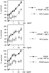
Dose–response curves of NT-3, IGF-2, and PDGF-BB alone and on the background of the other two factors. Note that each of these factors unambiguously promotes Schwann cell survival in a dose-dependent manner provided the other two factors are present. Note that when applied singly at the concentration used in the minimal mixture (IGF-2, 1.6 ng/ml; NT-3 and PDGF-BB, 0.8 ng/ml), the effect of each factor is small, although the mixture supports full survival (see Results and Fig. 2B, first column from left) showing a synergistic action in promoting survival. The IGF-2 curve was constructed in the presence of PDGF-BB and NT-3 both at 0.2 ng/ml. The NT-3 curve was constructed in the presence of PDGF-BB at 0.2 ng/ml and IGF-2 at 0.4 ng/ml. The PDGF-BB curve was constructed in the presence of NT-3 at 0.2 ng/ml and IGF-2 at 0.4 ng/ml. PORN substrate, 125 cells per coverslip, 2 d assay.
Fig. 4.
Fig. 4.
β-Neuregulins support Schwann cell survival but are unlikely to be a major component of the survival signal in conditioned media. A, A soluble form of the β-neuregulin-1 receptor ErbB4 inhibits β-neuregulin-1-mediated survival but has no effect on survival mediated by the minimal mixture of IGF-2, NT-3, and PDGF-BB or conditioned medium (dilution, 1:10). Note that for direct comparison, cell number at the end of the assay (2 d) in β-neuregulin-1 alone is arbitrarily set at 100%. Cell number in the presence of β-neuregulin-1 plus added ErbB4 is normalized to this value to obtain relative survival.NRG1, β-Neuregulin-1, 8 ng/ml. PORN substrate, 125 cells per coverslip, 2 d assay. B, β-Neuregulin-mediated survival is unaffected by neutralizing antibodies to IGF, NT-3, or PDGF-BB. DM, Simple defined medium; NRG1, β-neuregulin-1, 8 ng/ml. For anti-NT-3′ and anti-NT-3" see legend to Figure 2A; anti-IGF, IGF antibody SM1.2. The antibodies were applied at the same concentrations as those used to inhibit activity in conditioned media. PORN substrate, 125 cells per coverslip, 2 d assay.
Fig. 5.
Fig. 5.
Survival in conditioned medium or in IGF-2, NT-3, and PDGF-BB depends on MAP kinase activation, but survival in β-neuregulin-1 is MAP kinase-independent. The graph shows the effect of blocking the MAP kinase pathway (using 20, 35, or 50 μm PD98059) on survival under three different conditions: in the minimal mixture of IGF-2, NT-3, and PDGF-BB, in conditioned medium at a dilution of 1:10 (Cond.medium), and in β-neuregulin-1 at 8 ng/ml (NRG1). Note that PD98059 has no effect on cell numbers in β-neuregulin. The number is higher at the end of the assay than the number of cells initially plated because of the mitogenic effect of β-neuregulin-1. The experiment was done in simple defined medium using PORN substrate, 125 cells per coverslip, and a 2 d assay.
Fig. 6.
Fig. 6.
Semiquantitative RT-PCR analysis of the putative autocrine factors and their respective receptor mRNAs during development, in culture, and after nerve transection. A, RT-PCR on neonatal sciatic nerve (Ne) and Schwann cells cultured from neonatal nerves (Sch). Examination of IGF-1 (43 cycles), IGF-2 (28 cycles), PDGF-B (33 cycles), and NT-3 (48 cycles) and their corresponding receptors IGF-RI (45 cycles), IGF-RII (33 cycles), PDGF-Rβ (33 cycles), and TrkC (36 cycles) reveals the presence of all mRNAs in newborn nerve as well as Schwann cells in culture. B, These mRNAs are expressed in E18 nerves (immature Schwann cells) but also earlier in embryonic development at E14 (precursor stage) and E16 (a transition point between precursors and immature Schwann cells). The cycle numbers for each primer pair were 33 cycles for IGF-1, 27 cycles for IGF-2, 46 cycles for NT-3, and 33 cycles for the TrkC receptor. For all other primer pairs the cycle numbers were identical to those listed in A.C, After nerve transection, mRNAs for all factors and their receptors are expressed at the levels that are broadly comparable to the contralateral control side. CON, Normal nerves of 2- and 4-d-old rats; CUT, the distal stump of 2- and 4-d-old rats 2 and 4 d after transection performed at birth. Cycle numbers were 30 for IGF-1, 27 for IGF-2, 40 for NT-3, and 35 for TrkC. For PDGF-B and PDGF-Rβ 34 cycles were done. Again, 45 cycles were run for IGF-RI, and 33 were run for IGF-RII.
Fig. 7.
Fig. 7.
Immunohistochemical experiments on teased nerves and cultured Schwann cells. AC,E, G, I, Teased nerves from 4-week-old rats labeled by antibodies as indicated. The nerve shown in B is the distal stump of a nerve that was transected 3 d before immunolabeling. Note that labeling is more intense and widespread in B than in A. In myelinated fibers, immunolabeling is generally most intense in nuclear regions (examples indicated with double arrowheads) but is also seen in the outer cytoplasmic collar; this can be seen most clearly in E, G, and I. Examples of unmyelinated fibers are indicated with single arrowheads. D, F,H, J, Cultured Schwann cells made from 7-d-old rats. In all cases speckled immunolabeling was seen in the cell body region and in cellular processes. Scale bars (A,D), 20 μm.
Fig. 8.
Fig. 8.
In the presence of autocrine signals, laminin promotes long-term survival. A, In sparse cultures, Schwann cells die on laminin substrate. Laminin substrate, 125 cells per coverslip. B, Laminin promotes longer-term survival in the minimal mixture of IGF-2, NT-3, and PDGF-BB and supports full survival for at least 6 d in the presence of conditioned medium (CM; dilution, 1:10). Laminin substrate, 125 cells per coverslip.
Fig. 9.
Fig. 9.
The emergence of an autocrine survival circuit coincides with the generation of Schwann cells from precursors.A, Essentially all E14 precursors die irrespective of plating density, whereas the survival of immature Schwann cells from E18 nerves is density-dependent. Identical results were obtained on laminin or PORN substrates in experiments with precursors. E18 Schwann cells die more slowly on laminin than on PORN. Therefore, at the 2 d time points shown here, more cells are still alive at each density point on laminin compared with PORN substrates. One day assay for E14 precursors, 2 d assay for E18 Schwann cells. B, E14 Schwann cell precursors cannot be rescued by Schwann cell conditioned medium or by IGF-2, NT-3, and PDGF-BB. β-Neuregulin, an established precursor survival factor, is included as a positive control.DM, Simple defined medium; CM, Schwann cell conditioned medium at a dilution of 1:10. IGF-2, NT-3, and PDGF-BB were at the minimal concentrations, and β-neuregulin-1 was at 2 ng/ml. PORN substrate, 1 d assay. Identical results were obtained when the experiments shown in B were performed in supplemented defined medium and when the experiments were performed on laminin substrate (data not shown).
Fig. 10.
Fig. 10.
From paracrine signals to autocrine loops: the proposed changes in survival regulation during Schwann cell development. The survival of E14 Schwann cell precursors is regulated in a paracrine manner by axon-derived β-neuregulin-1. During development, Schwann cells establish autocrine circuits, which, in neonatal nerves, act in parallel with the axonal signal. During Schwann cell maturation the autocrine signal becomes sufficient to prevent Schwann cell death in the absence of axons. The factors IGF, NT-3, and PDGF-BB have been shown to be major components of this autocrine signal in Schwann cells from 7-d-old nerves. In vivoexperiments indicate that at this date loss of axonal contact after nerve transection no longer triggers significant Schwann cell death (Z. Dong, R. Mirsky, and K. R. Jessen, unpublished observations).A, Axons; P, Schwann cell precursors;S, Schwann cells.

Similar articles

Cited by

References

    1. Alessi DR, Cuenda A, Cohen P, Dudley DT, Saltiel AR. PD 098059 is a specific inhibitor of the activation of mitogen-activated protein kinase kinase in vitro and in vivo. J Biol Chem. 1995;270:27489–27494. - PubMed
    1. Barres BA, Hart IK, Coles HSR, Burne JF, Voyvodic JT, Richardson WD, Raff MC. Cell death and control of cell survival in the oligodendrocyte lineage. Cell. 1992;70:31–46. - PubMed
    1. Barres BA, Jacobson MD, Schmid R, Sendtner M, Raff MC. Does oligodendrocyte survival depend on axons? Curr Biol. 1993a;3:489–497. - PubMed
    1. Barres BA, Schmid R, Sendtner M, Raff MC. Multiple extracellular signals are required for long-term oligodendrocyte survival. Development. 1993b;118:283–295. - PubMed
    1. Bell GI, Stempien MM, Fong NM, Rall LB. Sequences of liver cDNAs encoding two different mouse insulin-like growth factor I precursors. Nucleic Acids Res. 1986;14:7873–7882. - PMC - PubMed

Publication types

MeSH terms

LinkOut - more resources