. 1999 May 15;19(10):3874-87.
doi: 10.1523/JNEUROSCI.19-10-03874.1999.

Spontaneous activity in developing turtle retinal ganglion cells: pharmacological studies

Affiliations

Spontaneous activity in developing turtle retinal ganglion cells: pharmacological studies

E Sernagor et al. J Neurosci. .

Abstract

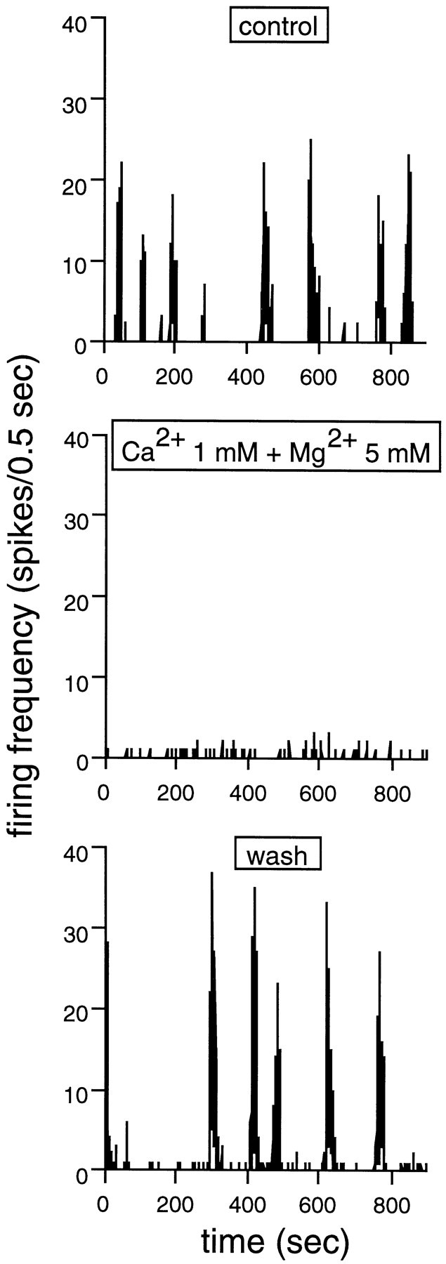
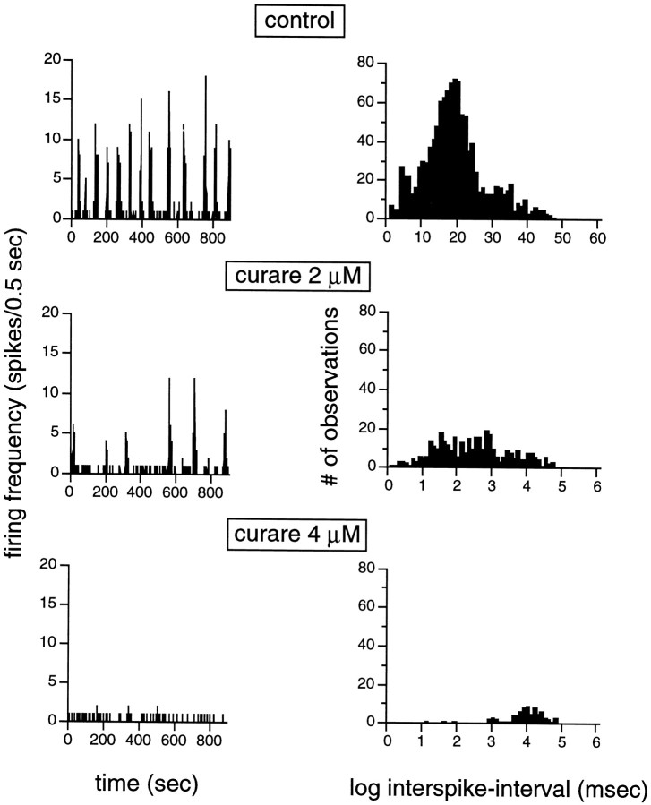
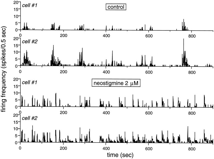
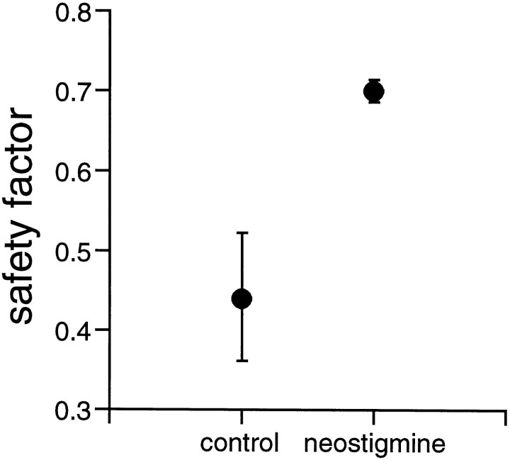
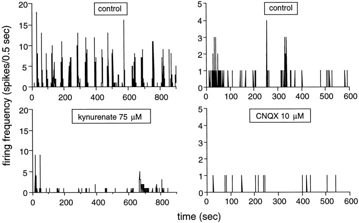
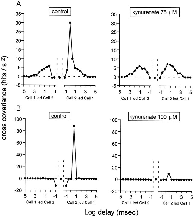
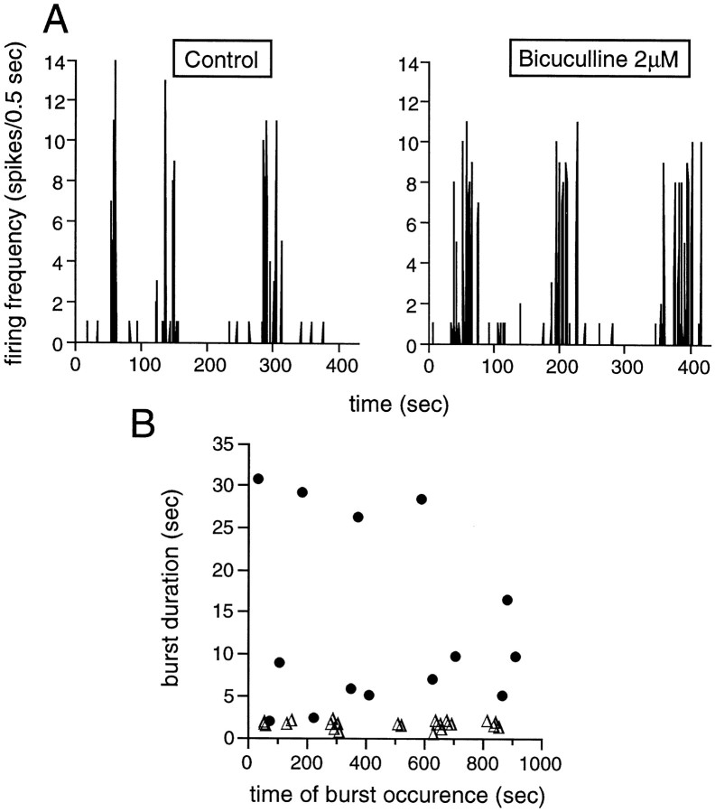
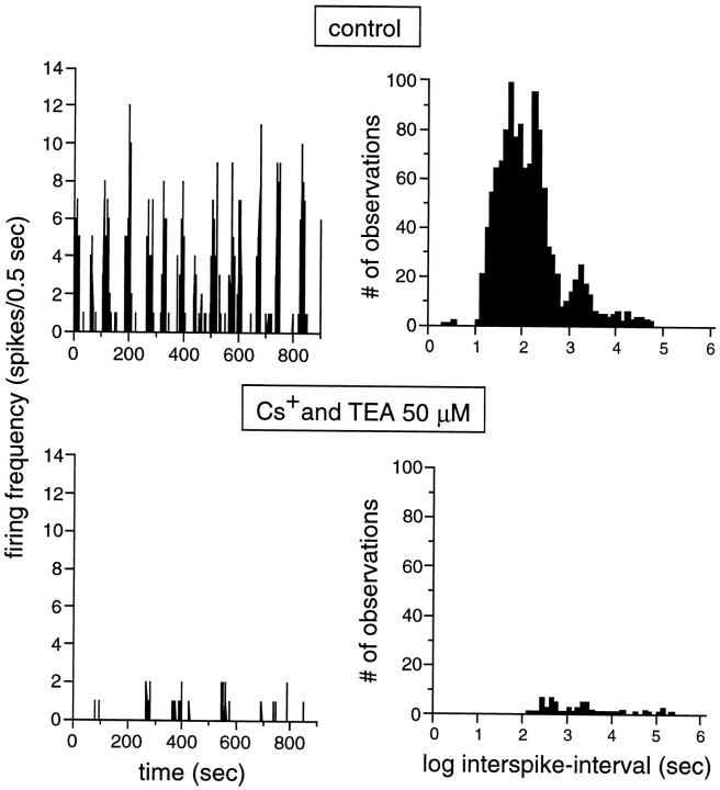
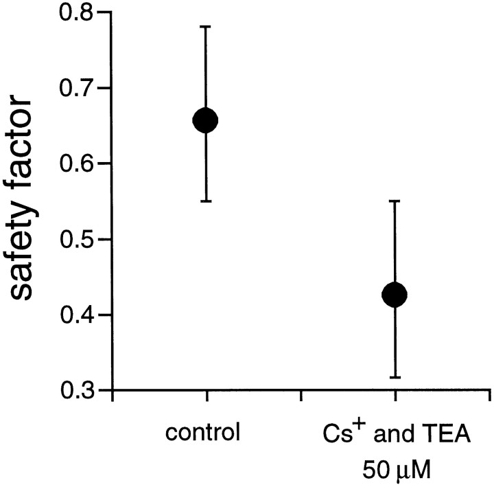
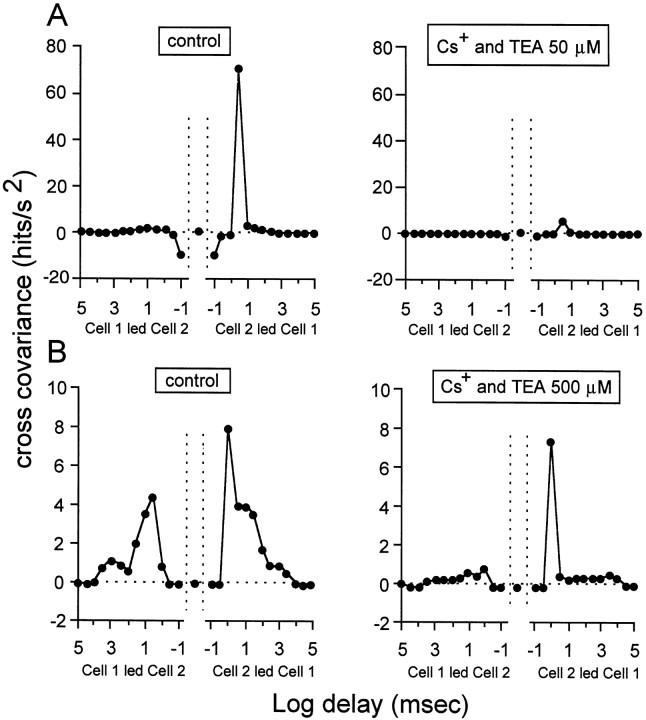
Extracellular recordings were obtained from the ganglion cell (GC) layer during correlated spontaneous bursting activity (SBA) in the immature turtle retina. Pharmacological agents were bath-applied, and their effects on burst and correlation parameters were determined. SBA requires synaptic transmission. It was blocked in the presence of curare and mecamylamine, two cholinergic nicotinic antagonists, and enhanced with neostigmine, a cholinesterase inhibitor. SBA was profoundly inhibited during blockade of glutamatergic receptors with the broad spectrum antagonist kynurenate and it vanished with 6,7-dinitroquinoxaline-2-3-dione (DNQX) and 6-cyano-7-nitroquinoxaline-2,3-dione (CNQX), two AMPA/kainate receptor antagonists. Blockade of NMDA receptors with D(-)-2-amino-5-phosphonopentanoic acid (D-AP-5) led only to a modest reduction in SBA. Blockade of GABAA receptors with bicuculline prolonged the duration of the bursts. Inhibition of GABA uptake with nipecotic acid led to a decrease in burst rate. Blockade of K+ channels with cesium (Cs+) and tetraethylammonium (TEA) led to a dramatic decrease in excitability. Burst propagation between neighboring GCs was reduced by K+ channel blockade. Gap junction blockade had no consistent effect on bursts or correlation parameters. None of these drugs had a strong effect on the refractory period between bursts. We conclude that correlated SBA in immature turtle GCs requires both cholinergic nicotinic and glutamatergic (mainly through AMPA/kainate receptors) synaptic transmission. GABAergic activity modulates the intensity and the duration of the bursts. Extracellular K+ is involved in lateral activity propagation and increases retinal excitability, which may be required for burst generation.


Figures

Fig. 1.
Fig. 1.
Synaptic transmission is required for spontaneous bursting. The spontaneous bursts are illustrated by changes in firing rate (in 0.5 sec bins) over a period of 900 sec. Burst onset and offset is indicated by abrupt changes in firing rate. Top panel, Control. The cell fires in strong, well defined bursts of spikes. Middle panel, When synaptic transmission is blocked by decreasing [Ca2+]out to 1 mm and increasing [Mg2+]out to 5 mm, the spontaneous bursts completely disappear. The only spontaneous activity left in these conditions is some isolated, sporadic spikes.Bottom panel, Wash. When [Ca2+]out and [Mg2+]out are returned to normal levels, strong spontaneous bursts reappear. Stage 25 GC.
Fig. 2.
Fig. 2.
Cholinergic nicotinic activity is required for the generation of spontaneous bursting activity. This figure illustrates how spontaneous bursts gradually disappear (both BR and FR within bursts decrease before the bursts disappear) as the concentration of curare, a cholinergic nicotinic blocker, increases from 2 to 4 μm. Top panels, Control. Middle panels, Activity in the presence of 2 μm curare.Bottom panels, Activity in the presence of 4 μm curare. Left panels, The spontaneous bursts are illustrated by changes in firing rate (in 0.5 sec bins) over a period of 900 sec, such as in Figure 1. Only isolated spontaneous spikes remain while the cholinergic activity is blocked. Right panels, Histograms illustrating interspike intervals (in logarithmic units) measured under different experimental conditions. As the curare concentration increases, the activity is slowed down, resulting in a shift to the right in the interspike intervals. In themiddle histogram, values represents those of the intervals between isolated spontaneous spikes. Stage 25 GC.
Fig. 3.
Fig. 3.
Spontaneous bursting activity is enhanced during blockade of cholinesterase with neostigmine. SBA is illustrated as in previous figures. The two top traces show the control activity in two adjacent GCs recorded by the same electrode.Cell #2 has stronger bursts and also more “background” activity superimposed on the bursts. The bursts are well synchronized between these two cells. The two bottom traces show the SBA in the presence of 2 μmneostigmine. Bursts are much more frequent. The firing rate within bursts has significantly increased in cell #1. The bursts are much shorter than before enhancement of acetylcholine activity by the drug. The activity is highly synchronized between the cells (see Fig. 4). Stage 25 GC.
Fig. 4.
Fig. 4.
The safety factor for activity propagation between adjacent cells increases during blockade of acetylcholinesterase with neostigmine. The figure illustrates the median SF for two pairs of cells in control conditions and in the presence of 2 μmneostigmine. When acetylcholinesterase is inhibited, the SF increases by 61.8%. Median absolute deviation bars. Stage 25 GCs.
Fig. 5.
Fig. 5.
Glutamatergic activity, acting mainly through AMPA/kainate receptors, is required for the generation of spontaneous bursting activity. SBA is illustrated as in previous figures. Theleft panels illustrate how kynurenate, a broad spectrum glutamate antagonist, profoundly reduces SBA. Top left panel, SBA in control conditions (S25 GC). The cell fires robust, regular bursts of spikes. There is very little background, sporadic activity in this cell. Bottom left panel, SBA in the presence of 75 μm kynurenate, a broad-spectrum antagonist of glutamatergic receptors. BR is significantly reduced in these conditions. The right panels illustrate that SBA disappears in the presence of CNQX, an AMPA/kainate receptor antagonist. Top right panel, SBA in control conditions (S26 GC). Bottom right panel, SBA in the presence of 10 μm CNQX. SBA completely vanishes in these conditions, leaving only few sporadic spikes. The disappearance of light responses was used in both experiments to demonstrate effective glutamate receptor blockade.
Fig. 6.
Fig. 6.
During spontaneous correlated bursting, glutamatergic activity appears to contribute to activity synchronization between neighboring cells by coordinating individual spikes between these cells. This figure illustrates the cross-covariance function for activity synchronization in two pairs of cells (A, B). The cross-covariance, expressed as hits per square second is plotted as a function of the delay in milliseconds in logarithmic units. Each point represents cross-covariance values averaged in intervals beginning at the abscissa of the point and extending half log unit. In normal conditions, this function has two components, a fast (1–3 msec), asymmetric component, followed by a slow, symmetric component. A, Theleft panel shows the control cross-covariance function for one pair of GCs. The right panel illustrates the function for the same pair of cells in the presence of 75 μm kynurenate. The fast, asymmetric component disappears in these conditions, whereas the slow component is unchanged. We may therefore conclude that glutamatergic activity is important for that early component of the function, which reflects synchronization between individual spikes in neighboring cells. B, Theleft panel illustrates the control cross-covariance function for another pair of cells (notice the difference in scale with that of A). The right panel illustrates the function for the same pair of cells in the presence of 100 μm kynurenate. Both components of the function have nearly vanished in these conditions. Values between −100 and 100 μsec are delimited by the vertical dotted lines. Stage 25 GCs.
Fig. 7.
Fig. 7.
GABA modulates the strength of the spontaneous bursts through GABAA receptors. A, These panels illustrate SBA, represented as in previous figures. Theleft panel shows SBA in control conditions, and theright panel in the presence of 2 μmbicuculline, a GABAA receptor antagonist. The bursts are longer in the presence of bicuculline, whereas BR does not change.B, This graph shows the duration of all the bursts (same GC as in A) occurring during the recording trial in control conditions (open triangles) and in the presence of 2 μm bicuculline (filled circles). The bursts are significantly longer when the GABAergic activity is blocked. Stage 25 GC.
Fig. 8.
Fig. 8.
Blockade of K+ channels, and thus of K+ efflux during activity, abolishes spontaneous bursting. Top panels, Control. Bottom panels, Activity in the presence of 50 μmCs+ and TEA, two K+ channel blockers. Right panels, Histograms of interspike intervals (in logarithmic units). The strong spontaneous bursts recorded in control conditions vanish in the presence of the drugs. Only very few weak bursts remain in the presence of Cs+ and TEA. The distribution of interspike intervals shifts to higher values and loses its clear bimodality as bursts become hardly detectable. Stage 25 GC.
Fig. 9.
Fig. 9.
The safety factor for activity propagation between adjacent cells decreases during blockade of K+channels. The figure illustrates the median SF for two pairs of cells in control conditions and in the presence of 50 μmCs+ and TEA. When K+ channels are blocked, the SF decreases by 35.3%. Median absolute deviation bars. Stage 25 GCs.
Fig. 10.
Fig. 10.
During spontaneous correlated bursting, extracellular K+ appears to be important not only for burst generation, but also for burst propagation between neighboring cells. As in Figure 6, this figure illustrates the cross-covariance function for activity synchronization in two pairs of cells (A, B). A, Theleft panel shows the control cross-covariance function for one pair of GCs. The right panel illustrates the function for the same pair of cells in the presence of 50 μm Cs+ and TEA. Both components of the function disappear in these conditions, and SBA is reduced to few weak clusters of spikes. B, The left panelillustrates the control cross-covariance function for another pair of cells. The right panel illustrates the function for the same pair of cells in the presence of 500 μmCs+ and TEA. Spontaneous firing resumes (see Results for explanation) in these conditions. The late, symmetric component of the cross-covariance function disappears, whereas the early, asymmetric component is intact. This demonstrates that extracellular K+ is important for the symmetric burst propagation between neighboring cells. Same graph conventions as for Figure 6. Stage 25 GCs.

Similar articles

Cited by

References

    1. Ariel M, Adolph AR. Neurotransmitter inputs to directionally sensitive turtle retinal ganglion cells. J Neurophysiol. 1985;54:1123–1143. - PubMed
    1. Arnett D, Spraker TE. Cross-correlation analysis of the maintained discharge of rabbit retinal ganglion cells. J Physiol (Lond) 1981;317:29–47. - PMC - PubMed
    1. Backman SB, Stein RD, Blank DW, Collier B, Polosa C. Different properties of the bradycardia produced by neostigmine and edrophonium in the cat. Can J Anaesth. 1996;43:731–740. - PubMed
    1. Baldridge WH. Optical recording of the effects of cholinergic ligands on neurons in the ganglion cell layer of mammalian retina. J Neurosci. 1995;15:5060–5072. - PMC - PubMed
    1. Brivanlou IH, Warland DK, Meister M. Mechanisms of concerted firing among retinal ganglion cells. Neuron. 1998;20:527–539. - PubMed

Publication types

MeSH terms

Substances

LinkOut - more resources