

Genesis, neurotrophin responsiveness, and apoptosis of a pronounced direct connection between the two eyes of the chick embryo: a natural error or a meaningful developmental event?
- PMID: 10234021
- PMCID: PMC6782693
- DOI: 10.1523/JNEUROSCI.19-10-03900.1999
Genesis, neurotrophin responsiveness, and apoptosis of a pronounced direct connection between the two eyes of the chick embryo: a natural error or a meaningful developmental event?
Abstract
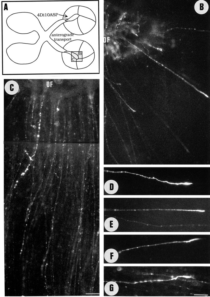
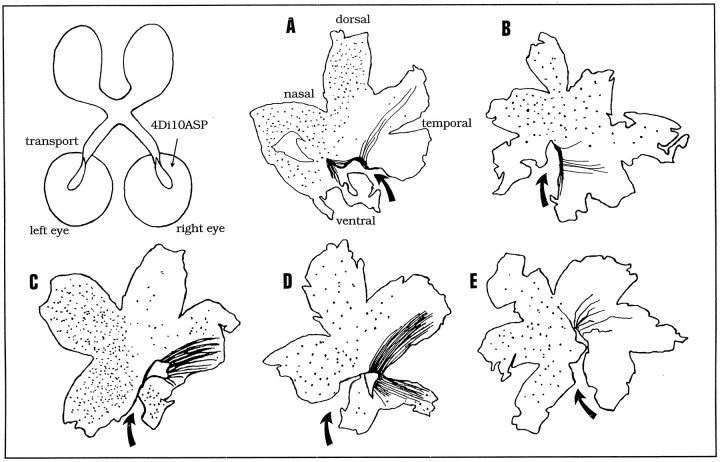
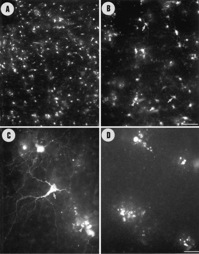
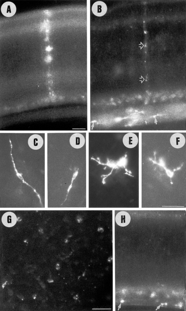
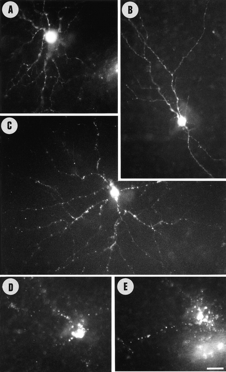
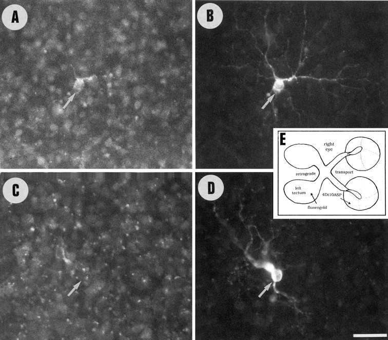
Unilateral intraocular injections of either of two fluorescent carbocyanine dyes into the embryonic chick eye resulted in both retrograde staining of ganglion cells (GCs) in the eye contralateral to site of injection and anterograde labeling of axons whose cell bodies were located within the injected eye. This prominent retino-retinal projection formed by thousands of GCs having a nasal origin and temporal termination appeared at embryonic day 6 (E6), attained its maximum intensity at E13-E14, and gradually disappeared until E18. The axonal growth cones ended superficially and never penetrated deeper layers of the retina. Treatment of the projection with BDNF resulted in massive terminal branching of the axons within deeper layers of the target retina. Double injection into the eye and the isthmo-optic nucleus showed a concomitant ingrowth of axons in the contralateral retina. Individual GCs died between E9 and E13, but massive apoptotic cell death was mainly monitored at E14 and later. Disintegrated cells showed typical images of apoptosis. Because degenerating cells were prelabeled with the membranophilic fluorescent carbocyanine dye, their death allowed the concomitant visualization of phagocytosing cells, too. Radial Müller glia were the only class of cells observed to become phagocytotic between E9 and E16. These cells became replaced exclusively with microglial cells from E17 on. The results suggest that the topologically restricted retino-retinal projection may have some developmental significance rather than representing a massive erroneous projection. Most likely, the projection may serve as a "template" to guide centrifugal isthmo-optic axons into the retina.
Figures


References
-
- Alvarez-Bolado G, Schwarz M, Gruss P. PAX-2 in the chiasm. Cell Tissue Res. 1997;290:197–200. - PubMed
-
- Apkarian P, Eckhardt PG, van-Schooneveld MJ. Detection of optic pathway misrouting in the human albino neonate. Neuropediatrics. 1991;22:211–215. - PubMed
-
- Bohn RC, Stelzner DJ. The aberrant retino-retinal projection during optic nerve regeneration in the frog. I. Time course of formation and cells of origin. J Comp Neurol. 1981a;196:605–620. - PubMed
-
- Bohn RC, Stelzner DJ. The aberrant retino-retinal projection during optic nerve regeneration in the frog. II. Anterograde labelling with horseradish peroxidase. J Comp Neurol. 1981b;196:621–632. - PubMed
Publication types
MeSH terms
Substances
LinkOut - more resources
Full Text Sources