Comparative Study
. 1999 May 15;19(10):3982-91.
doi: 10.1523/JNEUROSCI.19-10-03982.1999.

Differential regulation of the expression of corticotropin-releasing factor receptor type 2 (CRF2) in hypothalamus and amygdala of the immature rat by sensory input and food intake

Affiliations
Comparative Study

Differential regulation of the expression of corticotropin-releasing factor receptor type 2 (CRF2) in hypothalamus and amygdala of the immature rat by sensory input and food intake

M Eghbal-Ahmadi et al. J Neurosci. .

Abstract

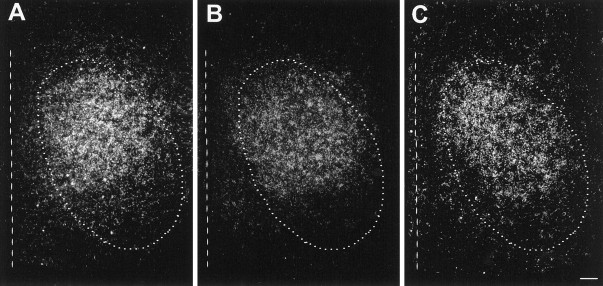
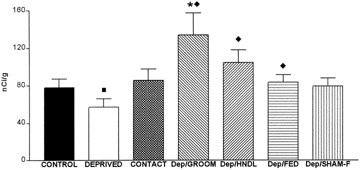
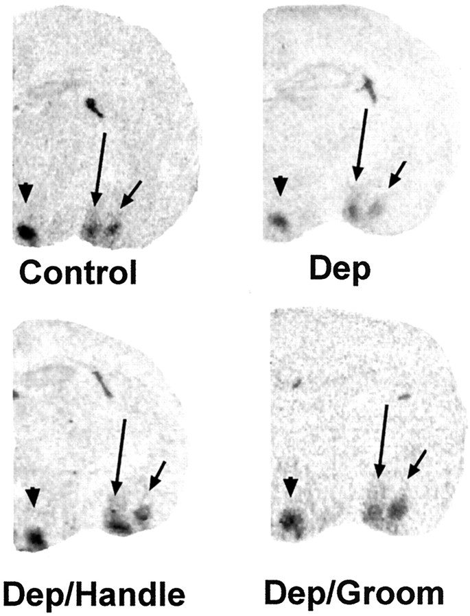
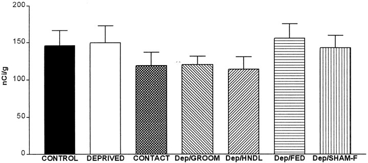
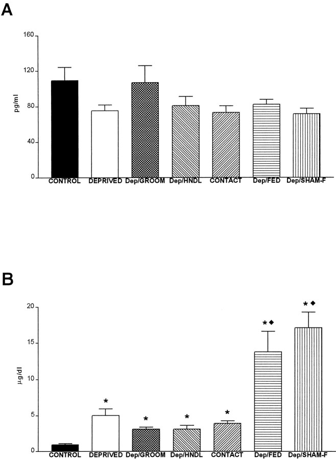
The physiological consequences of activating corticotropin-releasing factor receptor type 2 (CRF2) are not fully understood. The neuroanatomic distribution of this CRF receptor family member is consistent with roles in mediating the actions of CRF and similar ligands on food intake control and integrative aspects of stress-related behaviors. However, CRF2 expression in the adult rat is not influenced by stress, corticosterone (CORT), or food intake. In immature rat we have demonstrated striking downregulation of CRF2mRNA in hypothalamic ventromedial nucleus (VMH) after 24 hr of maternal deprivation, a paradigm consisting of both physiological/psychological stress and food deprivation. The current study aimed to distinguish which element or elements of maternal deprivation govern CRF2mRNA expression by isolating the effects of food intake and discrete maternal sensory cues on CRF2mRNA levels in VMH and in reciprocally communicating amygdala nuclei. In maternally deprived pups, CRF2mRNA levels in VMH and basomedial (BMA) and medial (MEA) amygdala nuclei were 62, 72, and 102% of control levels, respectively. Sensory inputs of grooming and handling as well as of the pups' own suckling activity-but not food intake-fully restored CRF2mRNA expression in VMH. In contrast, all manipulations tended to increase CRF2mRNA levels in BMA of maternally deprived rats, and surrogate grooming increased CRF2mRNA expression significantly above that of nondeprived controls. CRF2mRNA expression was not influenced significantly by plasma adrenocorticotropic hormone (ACTH) and CORT levels. Thus, in the immature rat, (1) CRF2 expression is regulated differentially in hypothalamic and amygdala regions, and (2) CRF2mRNA levels in VMH are governed primarily by maternal or suckling-derived sensory input rather than food intake or peripheral stress hormones. These findings indicate a region-specific regulation of CRF2mRNA, supporting the participation of the receptor in neurochemically defined circuits integrating sensory cues to influence specific behavioral and visceral functions.


Figures

Fig. 1.
Fig. 1.
CRF2mRNA expression in the VMH of immature rats subjected to selective physiological manipulations. A quantitative analysis of signal over VMH was achieved after ISH of matched coronal brain sections. Control, Animals were kept undisturbed in home cages; Deprived, pups were separated from the mother for 24 hr; Contact, pups were permitted contact with an anesthetized dam; Dep/Groom, maternally deprived animals were offered surrogate maternal grooming;Dep/HNDL, a deprived group was handled by a human as a control for the grooming; Dep/Fed, animals were gavage-fed an appropriate volume and calories of a milk-based diet;Dep/Sham-F, maternally deprived animals were undergoing the feeding procedure, but not receiving milk. *p< 0.05, significant difference from control; ♦ indicates significant difference from the maternally deprived group value. Values are expressed as means ± SEM. Group sizes (n) are found in Table 1.
Fig. 2.
Fig. 2.
Effect of physiological manipulations on CRF2mRNA levels in VMH of immature rats. Dark-field photomicrographs of coronal brain sections at the level of the VMH (encircled by the dotted line) after ISH that used a riboprobe directed against CRF2mRNA. Shown are a control rat (A), a rat after a 24 hr maternal deprivation (B), and a maternally deprived rat subjected to surrogate grooming (C). For orientation purposes the position of the third ventricle is indicated by a vertical dashed line. Note the localization of CRF2mRNA signal primarily over the dorsomedial portion of VMH. Scale bar, 70 μm.
Fig. 3.
Fig. 3.
CRF2mRNA expression in BMA of immature rats subjected to selective physiological manipulations. Quantitative analysis of signal over the BMA was achieved after ISH of matched coronal brain sections. Control, Animals kept undisturbed in home cages; Deprived, pups separated from the dam for 24 hr; Contact, pups permitted contact with an anesthetized dam; Dep/Groom, maternally deprived animals offered surrogate grooming; Dep/Hndl, a deprived group handled by a human as a control for the grooming;Dep/Fed, animals gavage-fed an appropriate volume and calories of a milk-based diet; Dep/Sham-F, maternally deprived animals undergoing the feeding procedure, but not receiving milk. *p < 0.05, significant difference from control; ♦p < 0.05, significant difference from the maternally deprived group; •p = 11. Values are expressed as means ± SEM. Group size is found in Table1.
Fig. 4.
Fig. 4.
Differential regulation of CRF2mRNA expression in specific hypothalamic and amygdala regions by discrete sensory input. Shown are photomicrographs of coronal brain sections at the level of VMH (arrowhead), MEA (long arrow), and BMA (short arrow). Sections were subjected to ISH for CRF2mRNA. Decreased signal over VMH and BMA is apparent in sections from a maternally deprived rat (Dep) as compared with control levels. Surrogate grooming (Dep/Groom) or handling by a human (Dep/Handle) enhanced the CRF2mRNA signal over VMH and BMA. No effect of any of the experimental manipulations on CRF2mRNA expression in MEA is evident (see also Fig.5).
Fig. 5.
Fig. 5.
CRF2mRNA expression in MEA of immature rats subjected to physiological manipulations. Quantitative analysis of signal over MEA was achieved after ISH of matched coronal brain sections. Group name abbreviations are as in Figure 1. No significant differences among values were observed (p > 0.05, ANOVA). Values are expressed as means ± SEM. The animal number for each group is found in Table 1.
Fig. 6.
Fig. 6.
Plasma stress hormone levels in immature rats subjected to maternal deprivation, and selective reinstitution of maternal sensory cues and feeding. A, ACTH plasma levels did not differ among groups. B, Plasma CORT levels of all maternally deprived groups were elevated significantly as compared with controls (*p < 0.01). In addition, CORT levels in groups subjected to the feeding procedure were significantly higher than those of both control and maternally deprived groups (♦p < 0.01). See Figure 1 for abbreviations.
Fig. 7.
Fig. 7.
Schematic of a proposed neurochemically defined circuit, using CRF or similar ligand and neurotransmitter and CRF2 as the receptor. Quantitative changes in CRF2mRNA expression may lead to altered neurotransmission in this circuit, which is involved in the transduction and integration of somatosensory, gustatory, and visceral signals and the neuroendocrine stress response. Blue frames indicate CRF2mRNA expression. Red shading over a region indicates the presence of CRF-expressing neurons. Red arrows denote established CRF-containing pathways.CEA, Central amygdaloid nucleus–bed nucleus of the stria terminalis continuum; NTS, nucleus of the solitary tract; PBN, parabrachial nucleus; SCN, suprachiasmatic hypothalamic nucleus.

Similar articles

Cited by

References

    1. Avishai-Eliner S, Yi SJ, Newth CJ, Baram TZ. Effects of maternal and sibling deprivation on basal and stress-induced hypothalamic–pituitary–adrenal components in the infant rat. Neurosci Lett. 1995;192:49–52. - PMC - PubMed
    1. Avishai-Eliner S, Yi SJ, Baram TZ. Developmental profile of messenger RNA for the corticotropin-releasing hormone receptor in the rat limbic system. Dev Brain Res. 1996;91:159–163. - PMC - PubMed
    1. Baram TZ, Hatalski CG. Neuropeptide-mediated excitability: a key triggering mechanism for seizure generation in the developing brain. Trends Neurosci. 1998;21:471–476. - PMC - PubMed
    1. Baram TZ, Lerner SP. Ontogeny of corticotropin releasing hormone gene expression in rat hypothalamus—comparison with somatostatin. Int J Dev Neurosci. 1991;9:473–478. - PubMed
    1. Behan DP, Heinrichs SC, Troncoso JC, Liu XJ, Kawas CH, Ling N, De Souza EB. Displacement of corticotropin releasing factor from its binding protein as a possible treatment for Alzheimer’s disease. Nature. 1995;378:284–287. - PubMed

Publication types

Substances