Enhanced neurotrophin-induced axon growth in myelinated portions of the CNS in mice lacking the p75 neurotrophin receptor
- PMID: 10234043
- PMCID: PMC6782726
- DOI: 10.1523/JNEUROSCI.19-10-04155.1999
Enhanced neurotrophin-induced axon growth in myelinated portions of the CNS in mice lacking the p75 neurotrophin receptor
Abstract
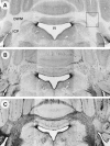
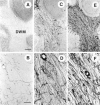
Axonal growth in the adult mammalian CNS is limited because of inhibitory influences of the glial environment and/or a lack of growth-promoting molecules. Here, we investigate whether supplementation of nerve growth factor (NGF) to the CNS during postnatal development and into adulthood can support the growth of sympathetic axons within myelinated portions of the maturing brain. We have also asked whether p75(NTR) plays a role in this NGF-induced axon growth. To address these questions we used two lines of transgenic mice overexpressing NGF centrally, with or without functional expression of p75(NTR) (NGF/p75(+/+) and NGF/p75(-/-) mice, respectively). Sympathetic axons invade the myelinated portions of the cerebellum, beginning shortly before the second week of postnatal life, in both lines of NGF transgenic mice. Despite the presence of central myelin, these sympathetic axons continue to sprout and increase in density between postnatal days 14 and 100, resulting in a dense plexus of sympathetic fibers within this myelinated environment. Surprisingly, the growth response of sympathetic fibers into the cerebellar white matter of NGF/p75(-/-) mice is enhanced, such that both the density and extent of axon ingrowth are increased, compared with age-matched NGF/p75(+/+) mice. These dissimilar growth responses cannot be attributed to differences in cerebellar levels of NGF protein or sympathetic neuron numbers between NGF/p75(+/+) and NGF/p75(-/-) mice. Our data provide evidence demonstrating that growth factors are capable of overcoming the inhibitory influences of central myelin in the adult CNS and that neutralization of the p75(NTR) may further enhance this growth response.
Figures


Similar articles
-
Nerve growth factor-induced growth of sympathetic axons into the optic tract of mature mice is enhanced by an absence of p75NTR expression.J Neurobiol. 1999 Apr;39(1):51-66. J Neurobiol. 1999. PMID: 10213453
-
Absence of the p75 neurotrophin receptor alters the pattern of sympathosensory sprouting in the trigeminal ganglia of mice overexpressing nerve growth factor.J Neurosci. 1999 Jan 1;19(1):258-73. doi: 10.1523/JNEUROSCI.19-01-00258.1999. J Neurosci. 1999. PMID: 9870956 Free PMC article.
-
Sympathetic and sensory axons invade the brains of nerve growth factor transgenic mice in the absence of p75NTR expression.Exp Neurol. 1998 Jan;149(1):284-94. doi: 10.1006/exnr.1997.6664. Exp Neurol. 1998. PMID: 9454638
-
Nerve Growth Factor in Alcohol Use Disorders.Curr Neuropharmacol. 2021;19(1):45-60. doi: 10.2174/1570159X18666200429003239. Curr Neuropharmacol. 2021. PMID: 32348226 Free PMC article. Review.
-
NGF in Neuropathic Pain: Understanding Its Role and Therapeutic Opportunities.Curr Issues Mol Biol. 2025 Jan 31;47(2):93. doi: 10.3390/cimb47020093. Curr Issues Mol Biol. 2025. PMID: 39996814 Free PMC article. Review.
Cited by
-
Glial inhibition of CNS axon regeneration.Nat Rev Neurosci. 2006 Aug;7(8):617-27. doi: 10.1038/nrn1956. Nat Rev Neurosci. 2006. PMID: 16858390 Free PMC article. Review.
-
ProNGF promotes neurite growth from a subset of NGF-dependent neurons by a p75NTR-dependent mechanism.Development. 2013 May;140(10):2108-17. doi: 10.1242/dev.085266. Development. 2013. PMID: 23633509 Free PMC article.
-
Neurotrophic factors and their receptors in axonal regeneration and functional recovery after peripheral nerve injury.Mol Neurobiol. 2003 Jun;27(3):277-324. doi: 10.1385/MN:27:3:277. Mol Neurobiol. 2003. PMID: 12845152 Review.
-
p75NTR-dependent, myelin-mediated axonal degeneration regulates neural connectivity in the adult brain.Nat Neurosci. 2010 May;13(5):559-66. doi: 10.1038/nn.2513. Epub 2010 Mar 28. Nat Neurosci. 2010. PMID: 20348920
-
Intracerebroventricular administration of α-ketoisocaproic acid decreases brain-derived neurotrophic factor and nerve growth factor levels in brain of young rats.Metab Brain Dis. 2016 Apr;31(2):377-83. doi: 10.1007/s11011-015-9768-8. Epub 2015 Nov 20. Metab Brain Dis. 2016. PMID: 26586008
References
-
- Barker PA, Shooter EM. Disruption of NGF binding to the low affinity neurotrophin receptor p75LNTR reduces NGF binding to TrkA on PC12 cells. Neuron. 1994;13:203–215. - PubMed
-
- Bregman BS, Kunkel-Bagden E, Schnell L, Dai HN, Gao D, Schwab ME. Regrowth of injured adult corticospinal and brainstem-spinal fibers elicited by antibodies to neurite growth inhibitors leads to recovery of locomotor function after spinal cord injury. Nature. 1995;378:498–501. - PubMed
-
- Bregman BS, McAtee M, Dai HN, Kuhn PL. Neurotrophic factors increase axonal growth after spinal cord injury and transplantation in the adult rat. Exp Neurol. 1997;148:475–494. - PubMed
-
- Caroni P, Schwab ME. Antibody against myelin-associated inhibitor of neurite growth neutralizes nonpermissive substrate properties of CNS white matter. Neuron. 1988a;1:85–96. - PubMed
Publication types
MeSH terms
Substances
Grants and funding
LinkOut - more resources
Full Text Sources
Other Literature Sources
Molecular Biology Databases
Research Materials
