

Anatomic fundoplication failure after laparoscopic antireflux surgery
- PMID: 10235525
- PMCID: PMC1420811
- DOI: 10.1097/00000658-199905000-00009
Anatomic fundoplication failure after laparoscopic antireflux surgery
Abstract
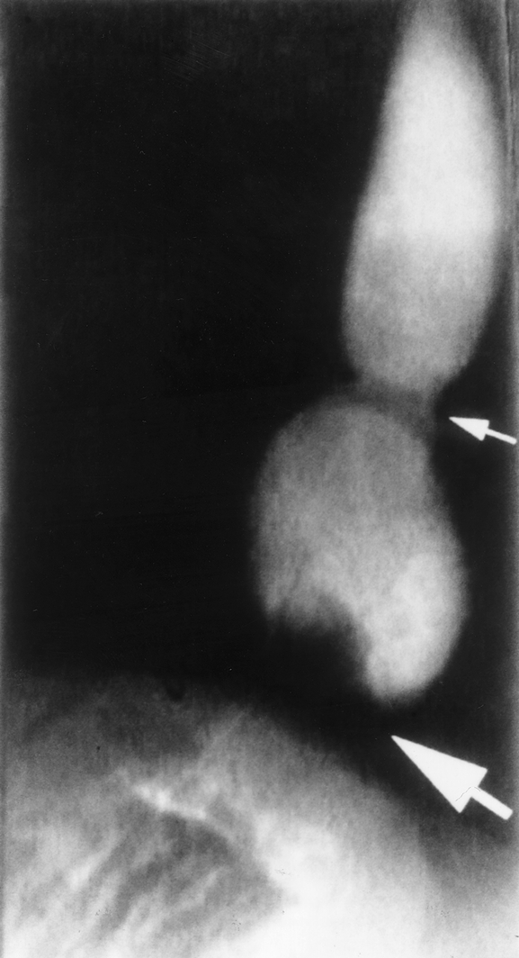
Objective: Anatomic fundoplication failure occurs after antireflux surgery and may be more common in the learning curve of laparoscopic antireflux surgery (LARS). The authors' aims were to assess the incidence, presentation, precipitating factors, and management of anatomic fundoplication failures after LARS.
Summary background data: The advent of LARS has increased the frequency with which antireflux surgery is performed for the treatment of gastroesophageal reflux disease. Postoperative symptoms frequently occur and may result from physiologic abnormalities or anatomic failure of the fundoplication (e.g., displacement or disruption). Few data exist on the potential causes or best treatment of anatomic fundoplication failures.
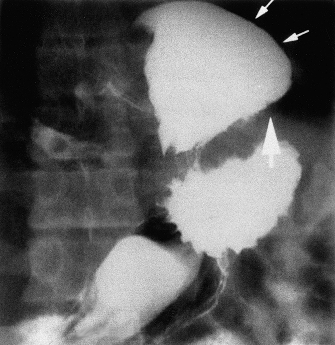
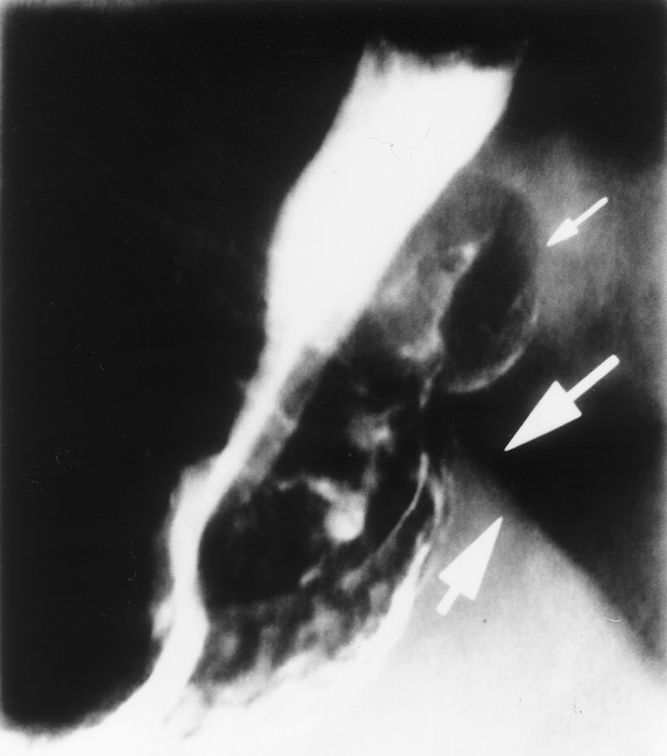
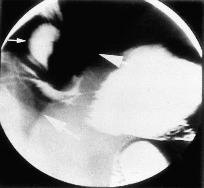
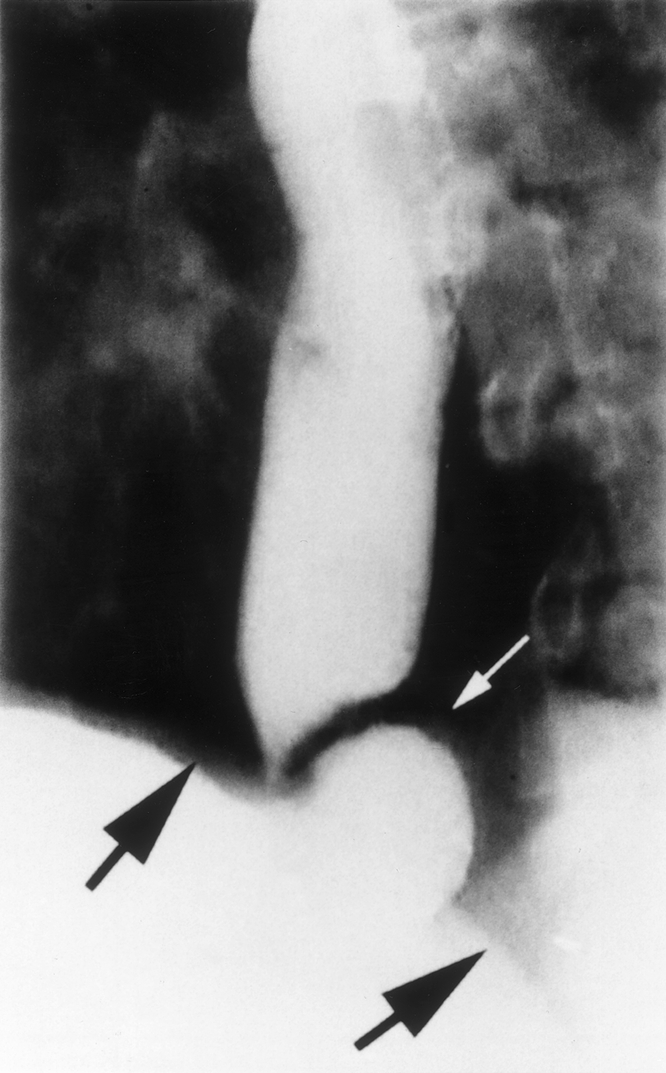
Method: LARS was performed in 290 patients by one of the authors over a 6-year period. In the first 53 patients (group 1), the short gastric vessels were divided on a selective basis and the diaphragmatic crura were closed only when large hiatal hernias were present. In the subsequent 237 patients (group 2), the crura were always approximated posterior to the short gastric vessels and full fundic mobilization was performed. Clinical postoperative evaluation was performed on a regular basis, with detailed tests of anatomy and physiology when untoward symptoms developed. Postoperative foregut symptoms were reported by 26% of the patients, of whom 73% were found to have an intact fundoplication. In 7% of the entire group, anatomic failure of the fundoplication was demonstrated, with the majority exhibiting intrathoracic migration of the wrap with or without disruption of the fundoplication. New-onset postoperative epigastric or substernal chest pain frequently heralded fundoplication failure. Factors correlated with the development of anatomic fundoplication failure included presence in group 1, early postoperative vomiting, other diaphragm "stressors," and large hiatal hernias. Repeat operation has been performed in 8 of the 20 patients (40%), with 5 patients successfully treated using laparoscopic techniques.
Conclusions: Anatomic fundoplication failure occurred in 7% of patients undergoing LARS, with the majority occurring in patients who underwent surgery during the learning curve. Anatomic failure is associated with technical shortcomings, large hiatal hernias, and early postoperative vomiting. Full esophageal mobilization and meticulous closure of the diaphragmatic crura posterior to the esophagus should minimize anatomic functional failure after LARS.
Figures
References
-
- Dallemagne B, Weerts JM, Jehaes C, et al. Laparoscopic Nissen fundoplication: preliminary report. Surg Laparosc Endosc 1991; 1: 138–143. - PubMed
-
- Perdikis G, Hinder RA, Lund RJ, et al. Laparoscopic Nissen fundoplication: where do we stand? Surg Laparosc Endosc 1997; 7: 17–21. - PubMed
-
- Laine S, Rantala A, Gullichsen R, Ovaska J. Laparoscopic vs. conventional Nissen fundoplication. Surg Endosc 1977; 11: 441–444. - PubMed
Publication types
MeSH terms
LinkOut - more resources
Full Text Sources
Medical
