. 1999 May 14;274(20):14382-91.
doi: 10.1074/jbc.274.20.14382.

A protein phosphatase methylesterase (PME-1) is one of several novel proteins stably associating with two inactive mutants of protein phosphatase 2A

Affiliations

A protein phosphatase methylesterase (PME-1) is one of several novel proteins stably associating with two inactive mutants of protein phosphatase 2A

E Ogris et al. J Biol Chem. .

Abstract

Carboxymethylation of proteins is a highly conserved means of regulation in eukaryotic cells. The protein phosphatase 2A (PP2A) catalytic (C) subunit is reversibly methylated at its carboxyl terminus by specific methyltransferase and methylesterase enzymes which have been purified, but not cloned. Carboxymethylation affects PP2A activity and varies during the cell cycle. Here, we report that substitution of glutamine for either of two putative active site histidines in the PP2A C subunit results in inactivation of PP2A and formation of stable complexes between PP2A and several cellular proteins. One of these cellular proteins, herein named protein phosphatase methylesterase-1 (PME-1), was purified and microsequenced, and its cDNA was cloned. PME-1 is conserved from yeast to human and contains a motif found in lipases having a catalytic triad-activated serine as their active site nucleophile. Bacterially expressed PME-1 demethylated PP2A C subunit in vitro, and okadaic acid, a known inhibitor of the PP2A methylesterase, inhibited this reaction. To our knowledge, PME-1 represents the first mammalian protein methylesterase to be cloned. Several lines of evidence indicate that, although there appears to be a role for C subunit carboxyl-terminal amino acids in PME-1 binding, amino acids other than those at the extreme carboxyl terminus of the C subunit also play an important role in PME-1 binding to a catalytically inactive mutant.


Figures
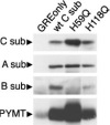
Fig. 1
Fig. 1. The catalytically inactive mutant PP2A C subunits can form complexes with A subunit and MT in vivo
Lysates from cells containing only control vector (GREonly) or HA-tagged wt (wt C sub) or mutant C subunits (H59Q and H118Q) were immunoprecipitated with anti-HA tag antibody (12CA5) and analyzed by SDS-PAGE and immunoblotting. The blot was probed first with anti-MT antibody, and then sequentially with antibodies recognizing the A, C (via the HA tag), and B PP2A subunits. Because a lower level of expression was consistently seen with H118Q, the immunoprecipitate of this mutant was prepared from more cells; to properly control for this, the control immunoprecipitate was prepared from an equivalent amount of cells expressing only the vector. Under these conditions, a small amount of MT can be seen sticking nonspecifically to the immunoprecipitate in the GREonly lane.
Fig. 2
Fig. 2. p44A appears to bind stoichiometrically to H118Q
Silver-stained two-dimensional gels of HA tag immunoprecipitates prepared from unlabeled cells expressing vector only (GREonly) or the C subunit mutant, H118Q, are shown. Only the portion of each gel containing the relevant proteins is shown. The A and C subunits, p44A, and anti-HA tag antibody heavy chain (Ab) are indicated by labeled brackets and arrowheads. Unlabeled arrowheads indicate the corresponding positions in the GREonly control panel. For reference, actin is indicated in both panels by a small unlabeled arrow. The approximate position where p44B would be located on these gels is indicated by the unlabeled brackets.
Fig. 3
Fig. 3. PME-1 cDNA schematic, mRNA tissue distribution, and predicted protein sequence
A, schematic of a 2.5-kilobase human PME-1 cDNA. On the stick diagram, the positions of the in-frame 5′-UTR stop codon (TGA), of the first two potential start codons (ATGs), of tandem stop codons (TAGTGA) at the end of the PME-1 ORF, and of the poly(A) tail (bracket) are shown. The 3′ end of the 3′-UTR, including the position of the poly(A) tail, was deduced by analyzing overlapping PME-1 ESTs; all other regions were directly sequenced. The sequence shown below the stick diagram extends from the in-frame 5′-UTR stop codon (TGA; overlined) to the second possible start ATG (double underlined). The first possible start ATG (underlined once in the sequence shown) was identified as the authentic start site in vivo by making constructs whose transcription/translation products in vitro would start with one or the other of these two ATGs. 35S-Labeled in vitro transcription/translation product starting at the first ATG, but not the product starting at the second ATG, comigrated precisely on two-dimensional gels with PME-1 from HeLa cell lysates (data not shown). B, expression of PME-1 mRNA in different tissues. Total RNA from the indicated mouse organs was separated by electrophoresis and hybridized with a mouse PME-1 partial cDNA probe from the 3′-UTR of mouse PME-1. In a separate experiment, the size of the PME-1 transcript was calculated to be 2.6 ± 0.2 kilobases. The lower panel shows the 18 S rRNA from the same blot visualized with methylene blue. C, the protein sequence encoded by the human PME-1 cDNA is shown. Amino acid sequence information for murine PME-1(p44A) obtained by tryptic peptide microsequencing is underlined. Over 98% of the microsequenced murine residues (107 of 109) were identical to the human sequence. The double underlined serine at position 42 corresponds to a threonine in murine PME-1. D, alignment of human, C. elegans (predicted), and S. cerevisiae (YHN5) PME-1 protein sequences. Residues identical with human PME-1 are shaded. Residues corresponding to the Prosite motif for lipases employing an active site serine are boxed.
Fig. 4
Fig. 4. PME-1 stably associates with H59Q but not wild-type C subunit
HA tag immunoprecipitates prepared from NIH3T3 (NIH) or MT-transformed NIH3T3 (NIHMT) cell lines individually expressing HA-tagged wt (wt C sub) or mutant (H59Q) C subunits were analyzed by SDS-PAGE and immunoblotting with HA tag antibody and PME-1 anti-peptide antibody. The C subunits migrate as tight doublets in these gels; whether doublets or a single band are seen varies from gel to gel and does not appear to be due to degradation (6, 13, 23). The panels and lanes shown are from the same experiment and gel, but the lanes were not all originally adjacent. Even on long exposure, the 44-kDa protein seen in the mutant lanes is not seen in the wt lanes.
Fig. 5
Fig. 5. Human PME-1 is a PP2A methylesterase
Immunoprecipitated PP2A C subunit was incubated with lysates from bacteria either not expressing PME-1 (control) or expressing PME-1 (PME-1), or with purified bacterially expressed PME-1 (~5 ng). Okadaic acid (O.A.) or PMSF was added to the reactions to the indicated final concentrations. Reactions containing 1.25% dimethyl sulfoxide (DMSO) as a control to match the level resulting from addition of okadaic acid or PMSF stock solutions are noted. After incubation, the immunoprecipitated PP2A C subunits were analyzed by SDS-PAGE. Proteins were transferred to nitrocellulose and the membrane was probed with 4b7 (methylation-sensitive Ab), an anti-C subunit antibody that only recognizes unmethylated C subunits. Subsequently, the same membrane was probed with Transduction Laboratories anti-PP2A C subunit antibody (methylation insensitive Ab), which is insensitive to the methylation state of PP2A and therefore reveals the total C subunit in each lane. The C subunits migrated as doublets in this gel, but whether double or single bands are seen can vary (see comments in legend to Fig. 4).
Fig. 6
Fig. 6. Analysis of H59Q·PME-1 complex formation
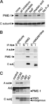
A, the PP2A inhibitors, okadaic acid, sodium fluoride, and sodium pyrophosphate, reduce the amount of PME-1 complexed with the catalytically inactive H59Q C subunit. Seven parallel dishes of NIH3T3 cells expressing HA-tagged H59Q were lysed as described under “Experimental Procedures” in Nonidet P-40 lysis buffer containing the indicated inhibitor(s) at the following concentrations: sodium vanadate (1 mm), sodim fluoride (50 mm), okadaic acid (500 nm), phenylarsine oxide (PAO, 10 μm), sodium pyrophosphate (NaP~P, 20 mm). Anti-HA tag immunoprecipitates were prepared from these lysates and analyzed by SDS-PAGE and immunoblotting. The blot was probed sequentially with antibodies detecting PME-1 and H59Q C subunit (via its HA tag). In a separate experiment using phosphorylase a as substrate (not shown), sodium fluoride, okadaic acid, and sodium pyrophosphate were, respectively, found to inhibit PP2A 91 ± 10, 97 ± 4, and >99%, while phenylarsine oxide and sodium vanadate, respectively, showed no or 25 ± 18% inhibition. B, loss of the C subunit carboxyl terminus reduces, but does not abolish, PME-1 binding. Non-immune (N) and HA tag (I) immuno-precipitates were prepared from MT-transformed NIH3T3 cells expressing vector only (GREonly), HA-tagged H59Q, or HA-tagged H59Q/301Stop double mutant which lacks nine carboxyl-terminal amino acids. Immune complexes were analyzed by SDS-PAGE; proteins were transferred to nitrocellulose; and immunoblotting was performed with antibodies directed against A subunit, PME-1, and C subunit (anti-HA tag). The C subunits migrate as doublets in this gel, but whether double or single bands are seen can vary (see comments in legend to Fig. 4). The band seen in all lanes in the PME-1 panel is from the immunoprecipitating antibodies. Chemiluminescent quantitation (using a Bio-Rad Fluor-S Max MultiImager or a Roche Molecular Biochemicals Lumiimager) was used in seven separate experiments with mixtures of clones to quantify the ratio of PME-1 to C subunit signal in each lane. In six of seven experiments with mixes of clones, the double mutant bound less PME-1 than did H59Q, with a mean reduction of 56 ± 30% and a median value of 39 (range of 8–87%). Thus, PME-1 binding is clearly reduced by loss of the carboxyl terminus. In a seventh experiment, for unknown reasons, the double mutant bound 235% of the H59Q level of PME-1, lowering the overall mean reduction to 28% (median = 40). C, C subunit carboxyl-terminal antibodies immunoprecipitate reduced amounts of H59Q·PME-1 complex. Immunoprecipitates were prepared from MT-transformed NIH3T3 cells expressing HA-tagged H59Q using control antibody, HA-tag antibody (12CA5), or carboxyl-terminal C subunit antibodies (1D6, 4B7, 4E1). The immune complexes were analyzed by SDS-PAGE, proteins were transferred to nitrocellulose, and immunoblotting was performed with anti-A subunit antibody (upper panel), anti-PME-1 antibody (middle panel), and anti-C subunit antibody recognizing both endogenous and HA tagged proteins (1D6 lower panel). The positions of A subunit, the immunoprecipitating antibody heavy chains (Ab), PME-1, HA-tagged H59Q C subunit, and untagged, endogenous wt C subunit are indicated. The C subunits migrate as single bands in this gel, but whether double or single bands are seen can vary (see comments in legend to Fig. 4). HA-tagged H59Q C subunit migrates more slowly than endogenous wt C subunit because of the HA tag.

Similar articles

Cited by

References

    1. Cohen P. Annu. Rev. Biochem. 1989;58:453–508. - PubMed
    1. Mumby MC, Walter G. Physiol. Rev. 1993;73:673–699. - PubMed
    1. Jakes S, Mellgren RL, Schlender KK. Biochim. Biophys. Acta. 1986;888:135–142. - PubMed
    1. Sim AT, Ratcliffe E, Mumby MC, Villa-Moruzzi E, Rostas JA. J. Neurochem. 1994;62:1552–1559. - PubMed
    1. Sontag E, Nunbhakdi-Craig V, Bloom GS, Mumby MC. J. Cell Biol. 1995;128:1131–1144. - PMC - PubMed

Publication types

MeSH terms

Associated data