. 1999 May 11;96(10):5486-91.
doi: 10.1073/pnas.96.10.5486.

Solution structure and dynamics of a de novo designed three-helix bundle protein

Affiliations

Solution structure and dynamics of a de novo designed three-helix bundle protein

S T Walsh et al. Proc Natl Acad Sci U S A. .

Abstract

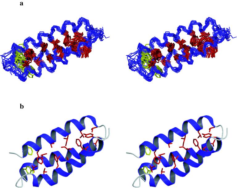
Although de novo protein design is an important endeavor with implications for understanding protein folding, until now, structures have been determined for only a few 25- to 30-residue designed miniproteins. Here, the NMR solution structure of a complex 73-residue three-helix bundle protein, alpha3D, is reported. The structure of alpha3D was not based on any natural protein, and yet it shows thermodynamic and spectroscopic properties typical of native proteins. A variety of features contribute to its unique structure, including electrostatics, the packing of a diverse set of hydrophobic side chains, and a loop that incorporates common capping motifs. Thus, it is now possible to design a complex protein with a well defined and predictable three-dimensional structure.


Figures

Figure 1
Figure 1
Sequences of the α3 family and of Coil-Ser. The residues are aligned with their corresponding heptad position in a coiled coil (26). α3A through α3C and Coil-Ser were chemically synthesized whereas α3D was cloned and expressed in E. coli. The residues that are different between α3D and α3C are labeled in bold.
Figure 2
Figure 2
(a) NMR spectrum of α3D (1H,13C-CT–heteronuclear single-quantum coherence) illustrating the assignments of the methyl groups. Prochiral assignments of the methyl groups of valine and leucine were obtained by using a 10% 13C labeled sample (35). (b) Summary of the α3D sequential NOEs. The size of the bar corresponds to the intensity of the NOE. The NOEs were taken from the 15N- and 13C-resolved nuclear Overhauser effect spectroscopy spectra. The three bond 3JαN coupling constants determined for α3D are displayed as filled circles and boxes corresponding to 3JαN coupling constants <6.0 Hz or >8.0 Hz, respectively.
Figure 3
Figure 3
(a) Stereo diagrams of the 13 superimposed α3D structures are shown with the hydrophobic core residues depicted in red and W4 and Y45 in yellow. The structures were aligned by using only the backbone atoms (residues 4–21, 24–45, and 51–70). The figure was generated by using the program molmol (58). (b) Stereo display of a ribbon diagram with the hydrophobic residues in red and W4 and Y45 in yellow of the lowest energy structure of α3D. The figure was created by using the programs molscript (59) and raster3d (60).
Figure 4
Figure 4
(a) Protection factors for the main-chain amide protons of α3D determined from hydrogen–deuterium exchange at 15°C, pH* 5.0 (in 50 mM deuterated acetate). The protection factors were calculated from the observed exchange rates and the intrinsic exchange rates (39). Absence of a bar indicates residues with amide protons exchanging in the dead time of the experiment (18 minutes) or a lack of a well resolved amide proton cross peak. (b) Assuming an EX2 exchange mechanism, the apparent free energy, ΔGHX, was calculated from the protection factors, Pi, as RTln(Pi) (39). The dashed line represents the ΔGu at 15°C determined calorimetrically by extrapolation of the Gibbs-Helmoltz equation, ΔGT = ΔHTm (1 − T/Tm) + (TTm) ΔCpTΔCpln(T/Tm). The shaded region denotes the estimated standard error in this parameter of ±0.5 kcal⋅mol−1.

Similar articles

Cited by

References

    1. Bryson J W, Betz S F, Lu H S, Suich D J, Zhou H X, O’Neil K T, DeGrado W F. Science. 1995;270:935–941. - PubMed
    1. Beasley J R, Hecht M H. J Biol Chem. 1997;272:2031–2034. - PubMed
    1. Hellinga H. Fold Des. 1998;3:1–8. - PubMed
    1. Regan L. Structure (London) 1998;6:1–4. - PubMed
    1. Choo Y, Castellanos A, Garcia-Hernandez B, Sanchez-Garcia I, Klug A. J Mol Biol. 1997;273:525–532. - PubMed

Publication types

Associated data

LinkOut - more resources