

The developing renal, reproductive, and respiratory systems of the African elephant suggest an aquatic ancestry
- PMID: 10318922
- PMCID: PMC21898
- DOI: 10.1073/pnas.96.10.5555
The developing renal, reproductive, and respiratory systems of the African elephant suggest an aquatic ancestry
Abstract
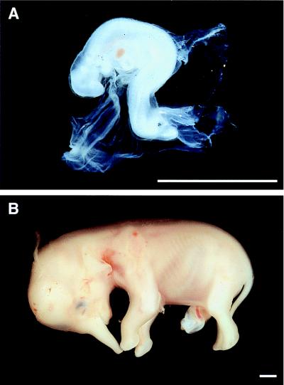
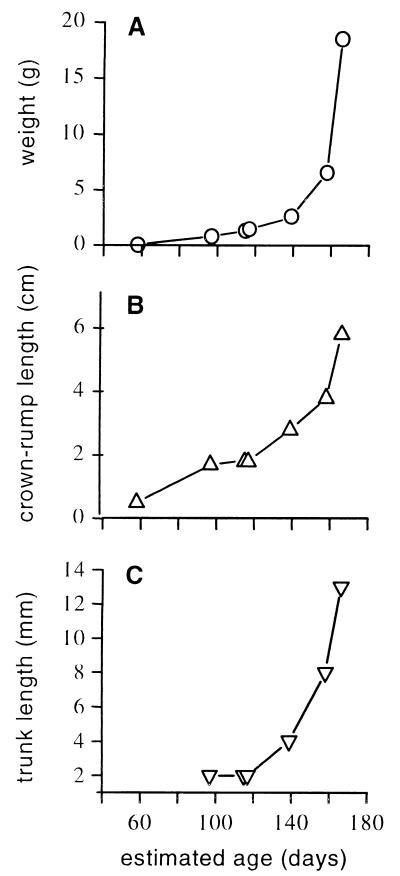
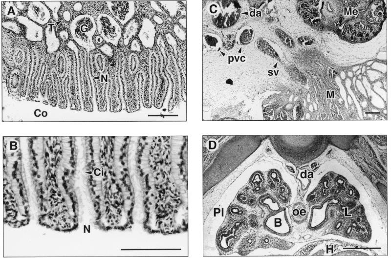
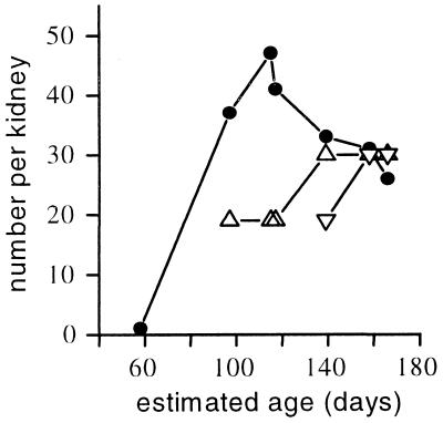
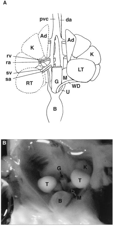
The early embryology of the elephant has never been studied before. We have obtained a rare series of African elephant (Loxodonta africana) embryos and fetuses ranging in weight from 0.04 to 18.5 g, estimated gestational ages 58-166 days (duration of gestation is approximately 660 days). Nephrostomes, a feature of aquatic vertebrates, were found in the mesonephric kidneys at all stages of development whereas they have never been recorded in the mesonephric kidneys of other viviparous mammals. The trunk was well developed even in the earliest fetus. The testes were intra-abdominal, and there was no evidence of a gubernaculum, pampiniform plexus, processus vaginalis, or a scrotum, confirming that the elephant, like the dugong, is one of the few primary testicond mammals. The palaeontological evidence suggests that the elephant's ancestors were aquatic, and recent immunological and molecular evidence shows an extremely close affinity between present-day elephants and the aquatic Sirenia (dugong and manatees). The evidence from our embryological study of the elephant also suggests that it evolved from an aquatic mammal.
Figures
Similar articles
-
Fetal lung development in the elephant reflects the adaptations required for snorkeling in adult life.Respir Physiol Neurobiol. 2003 Nov 14;138(2-3):325-33. doi: 10.1016/s1569-9048(03)00199-x. Respir Physiol Neurobiol. 2003. PMID: 14609520
-
Development of the germinal ridge and ovary in the African elephant (Loxodonta africana).Reproduction. 2012 Nov;144(5):583-93. doi: 10.1530/REP-12-0303. Epub 2012 Sep 18. Reproduction. 2012. PMID: 22991581
-
DETECTION OF ELEPHANT ENDOTHELIOTROPIC HERPESVIRUS (EEHV) IN FREE-RANGING AFRICAN ELEPHANTS (LOXODONTA AFRICANA) IN THE KRUGER NATIONAL PARK, SOUTH AFRICA.J Wildl Dis. 2023 Jan 1;59(1):128-137. doi: 10.7589/JWD-D-22-00015. J Wildl Dis. 2023. PMID: 36584337
-
The testicular descent in human. Origin, development and fate of the gubernaculum Hunteri, processus vaginalis peritonei, and gonadal ligaments.Adv Anat Embryol Cell Biol. 2000;156:III-X, 1-98. Adv Anat Embryol Cell Biol. 2000. PMID: 11008363 Review.
-
The chromosomes of Afrotheria and their bearing on mammalian genome evolution.Cytogenet Genome Res. 2012;137(2-4):144-53. doi: 10.1159/000341387. Epub 2012 Aug 3. Cytogenet Genome Res. 2012. PMID: 22868637 Review.
Cited by
-
Elephants develop wrinkles through both form and function.R Soc Open Sci. 2024 Oct 9;11(10):240851. doi: 10.1098/rsos.240851. eCollection 2024 Oct. R Soc Open Sci. 2024. PMID: 39386989 Free PMC article.
-
Reappraising the exteriorization of the mammalian testes through evolutionary physiology.Commun Integr Biol. 2019 Mar 20;12(1):38-54. doi: 10.1080/19420889.2019.1586047. eCollection 2019. Commun Integr Biol. 2019. PMID: 31143362 Free PMC article. Review.
-
The evolution of the discrete multirenculate kidney in mammals from ecological and molecular perspectives.Genome Biol Evol. 2023 May 9;15(5):evad075. doi: 10.1093/gbe/evad075. Online ahead of print. Genome Biol Evol. 2023. PMID: 37159529 Free PMC article.
-
Foetal age determination and development in elephants.Proc Biol Sci. 2007 Feb 7;274(1608):323-31. doi: 10.1098/rspb.2006.3738. Proc Biol Sci. 2007. PMID: 17164195 Free PMC article.
-
Hypothesis: gonadal temperature influences sex-specific imprinting.Front Genet. 2014 Aug 25;5:294. doi: 10.3389/fgene.2014.00294. eCollection 2014. Front Genet. 2014. PMID: 25202325 Free PMC article.
References
-
- Janus C M. TREE. 1988;3:291–297. - PubMed
-
- Shoshani T, Tassy P. The Proboscidea. New York: Oxford Univ. Press; 1996. p. 472.
-
- Domning D P, Ray C E, McKenna M C. Smithson Contrib Paleontol. 1986;59:1–56.
-
- de Jong W W. In: Macromolecular Sequences in Systematic and Evolutionary Biology. Goodman M E, editor. New York: Plenum; 1982. pp. 75–144.
-
- Fischer M S, Tassey P. In: Mammal Phylogeny. Szalay F S, Novacek M J, McKenna M C, editors. New York: Springer; 1993. pp. 217–234.
MeSH terms
LinkOut - more resources
Full Text Sources
