. 1999 May 11;96(10):5610-5.
doi: 10.1073/pnas.96.10.5610.

A cell surface mucin specifically expressed in the midgut of the malaria mosquito Anopheles gambiae

Affiliations

A cell surface mucin specifically expressed in the midgut of the malaria mosquito Anopheles gambiae

Z Shen et al. Proc Natl Acad Sci U S A. .

Abstract

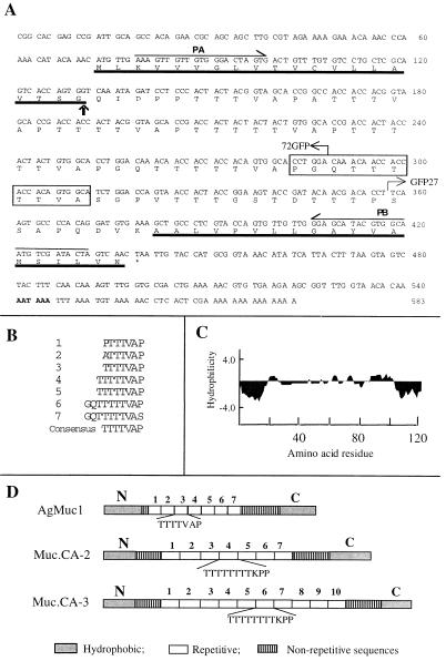
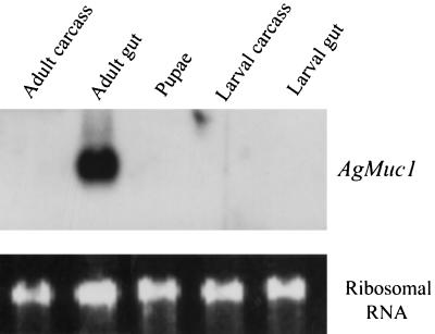
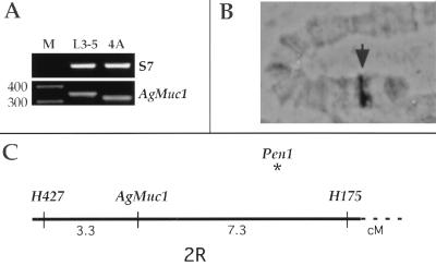
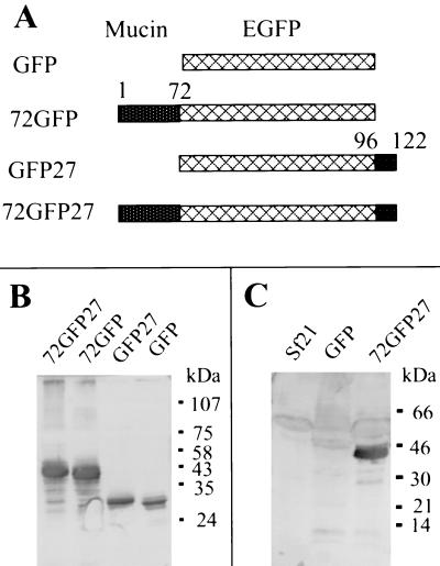
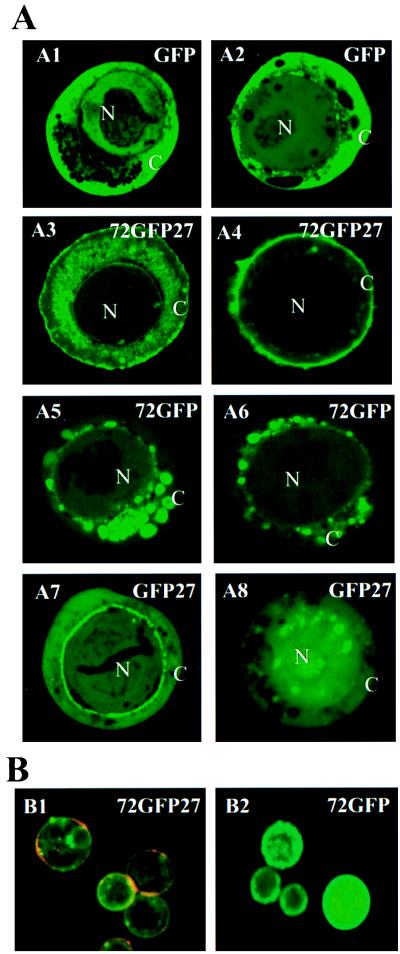
An invertebrate intestinal mucin gene, AgMuc1, was isolated from the malaria vector mosquito Anopheles gambiae. The predicted 122-residue protein consists of a central core of seven repeating TTTTVAP motifs flanked by hydrophobic N- and C-terminal domains. This structure is similar to that of mucins that coat the protozoan parasite Trypanosoma cruzi. Northern blot analysis indicated that the gene is expressed exclusively in the midgut of adult mosquitoes. A length polymorphism and in situ hybridization were used to genetically and cytogenetically map AgMuc1 to division 7A of the right arm of the second chromosome. The subcellular localization of the encoded protein in tissue culture cells was examined by using a baculovirus vector to express AgMuc1 protein tagged with the green fluorescent protein (GFP). The results indicated that this protein is found at the cell surface and that both hydrophobic domains are required for cell surface targeting. We propose that AgMuc1 is an abundant mucin-like protein that lines the surface of the midgut microvilli, potentially protecting the intestinal epithelium from the proteinase-rich environment of the gut lumen. An intriguing possibility is that, as an abundant surface protein, AgMuc1 may also interact with the malaria parasite during its invasion of the mosquito midgut.


Figures

Figure 1
Figure 1
Nucleotide and predicted amino acid sequences of AgMuc1. (A) The N- and C-terminal hydrophobic protein domains are underlined. The putative cleavage site of the signal peptide is indicated by a vertical arrow and the borders of GFP fusion constructs by bent arrows. The putative polyadenylation signal sequence is in boldface. The polymorphic region missing in the Muc1b allele is boxed and the PCR primers used for genetic mapping (PA, PB) are indicated above the sequence. (B) The seven repeated motifs of the central protein core are aligned. The consensus repeat sequence is given at the bottom. (C) A Kyte–Doolittle hydropathy plot was generated with an average hydrophilicity window of 7 residues. (D) Diagrammatic comparison of the predicted amino acid sequence of the A. gambiae AgMuc1 mucin with that of Trypanosoma cruzi mucins, MUC.CA-2 and MUC.CA-3 (31, 32). AgMuc1 mucin contains TTTTVAP repeats, whereas the MUC.CA-2 and MUC.CA-3 mucins contain TTTTTTTTKPP motifs. All three mucins contain nonrepeated sequences between the repeat array and the N- and C-terminal hydrophobic sequences.
Figure 2
Figure 2
Developmental and tissue specificity of AgMuc1 expression. (Upper) Autoradiogram of a Northern blot of RNAs (≈5 μg per lane) isolated from the indicated tissues. The blot was hybridized overnight with a 32P-labeled AgMuc1 cDNA probe, washed, and exposed to film for 4 h. (Lower) Staining of ribosomal RNA with ethidium bromide to indicate the amount of RNA analyzed in each lane.
Figure 3
Figure 3
Polymorphism and genetic mapping of AgMuc1. (A) Ethidium bromide-stained agarose gel with the PCR-amplified AgMuc1 segment containing the length polymorphism from the refractory L3-5 strain and susceptible 4A r/r strain. Lane M, length markers. (B) The cytogenetic location of AgMuc1 in division 7A on the second chromosome is indicated by an arrowhead. (C) Genetic distances of AgMuc1 from adjacent microsatellite markers are shown in centimorgans (cM). The gene is within the same region as the Plasmodium refractory QTL Pen1, but the latter locus is distinct from AgMuc1 and is located closer to H175.
Figure 4
Figure 4
Expression and characterization of mucin-GFP fusion proteins encoded by recombinant baculoviruses. (A) Diagrams showing recombinant protein structures. Lattice-patterned rectangles, GFP reporter protein; stippled rectangles, AgMuc1 sequences (see Fig. 1A). (B) Western blotting analysis of baculovirus-expressed GFP and GFP-mucin fusion proteins with an anti-GFP antibody. Total proteins from cells infected with different recombinant baculoviruses were loaded on each lane. A polyclonal antibody against GFP was used to probe the Western blots. The structure of the recombinant proteins indicated at the top of each lane is given in A. (C) Western blotting analysis of the baculovirus-expressed mucin-GFP protein with antiserum against total midgut microvilli. The Sf21 lane contains control cell lysate. Migration of marker proteins is indicated on the right side of B and C.
Figure 5
Figure 5
Localization of baculovirus-expressed GFP fusion proteins in Sf21 cells. (A) Sf21 cells were infected with recombinant baculoviruses that express the GFP, 72GFP27, GFP27, and 72GFP recombinant constructs (Fig. 4A) under the control of the viral polyhedrin promoter. Representative images recorded at 30 h after infection (A1, A3, A5, and A7) and at 60 h after infection (A2, A4, A6, and A8) are shown. The green is due to fluorescence of GFP or of GFP fusion proteins. N, Nucleus; C, cytoplasm. (×1000.) (B) Immunological localization of mucin-GFP fusion proteins. Living Sf21 cells expressing fusion proteins 72GFP27 or 72GFP (as shown in A4 and A6, respectively) were incubated with a rabbit anti-GFP antibody followed by incubation with a rhodamine-conjugated anti-rabbit IgG secondary antibody, thus detecting GFP-containing proteins only if exposed to the surface (red channel). The distribution of GFP, independent of its location in the cell, was detected by its inherent fluorescence (green channel). The images shown in B1 and B2 were collected while using identical settings for the green and red channels, and images from the red and green channels were then merged. (×250.)

References

    1. Billingsley P F, Lehane M J. In: Biology of the Insect Midgut. Lehane M J, Billingsley P F, editors. London: Chapman & Hall; 1996. pp. 3–25.
    1. Jacobs-Lorena M, Oo M M. In: The Biology of Disease Vectors. Beaty B J, Marquardt W C, editors. Niwot: Univ. Press of Colorado; 1996. pp. 318–332.
    1. Tellam R L. In: Biology of the Insect Midgut. Lehane M J, Billingsley P F, editors. London: Chapman & Hall; 1996. pp. 86–114.
    1. Del Bene G, Dallai R, Marchini D. Int J Morphol Embryol. 1991;20:15–24.
    1. Elvin C M, Vuocolo T, Pearson R D, East I J, Riding G A, Eisemann C H, Tellam R L. J Biol Chem. 1996;271:8925–8935. - PubMed

Publication types

MeSH terms

Associated data

LinkOut - more resources