. 1999 May 11;96(10):5651-6.
doi: 10.1073/pnas.96.10.5651.

Selection of functional T cell receptor mutants from a yeast surface-display library

Affiliations

Selection of functional T cell receptor mutants from a yeast surface-display library

M C Kieke et al. Proc Natl Acad Sci U S A. .

Abstract

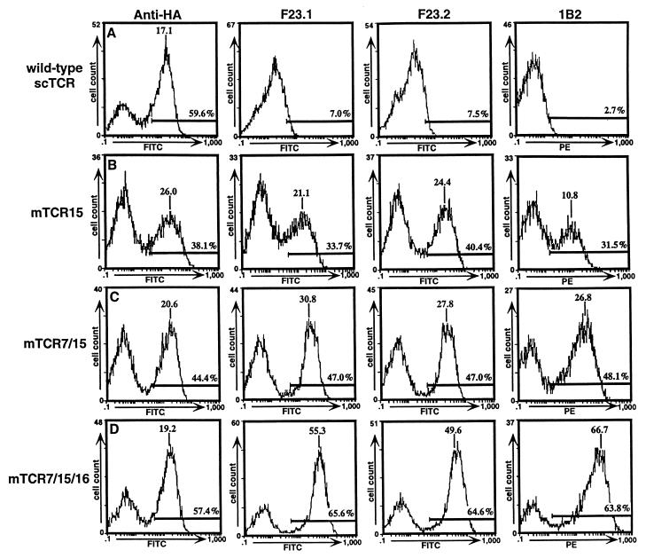
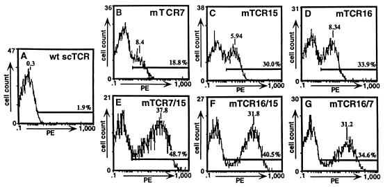
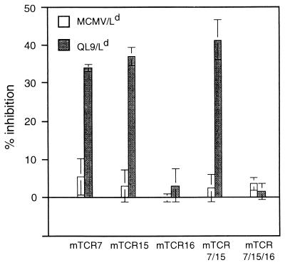
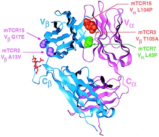
The heterodimeric alphabeta T cell receptor (TCR) for antigen is the key determinant of T cell specificity. The structure of the TCR is very similar to that of antibodies, but the engineering of TCRs by directed evolution with combinatorial display libraries has not been accomplished to date. Here, we report that yeast surface display of a TCR was achieved only after the mutation of specific variable region residues. These residues are located in two regions of the TCR, at the interface of the alpha- and beta-chains and in the beta-chain framework region that is thought to be in proximity to the CD3 signal-transduction complex. The mutations are encoded naturally in many antibody variable regions, indicating specific functional differences that have not been appreciated between TCRs and antibodies. The identification of these residues provides an explanation for the inherent difficulties in the display of wild-type TCRs compared with antibodies. Yeast-displayed mutant TCRs bind specifically to the peptide/MHC antigen, enabling engineering of soluble T cell receptors as specific T cell antagonists. This strategy of random mutagenesis followed by selection for surface expression may be of general use in the directed evolution of other eukaryotic proteins that are refractory to display.


Figures

Figure 1
Figure 1
Flow cytometric analysis of wild-type TCR/yeast and mutant TCR/yeast. Yeast displaying wild-type TCR (A), single-mutant mTCR15 (B), double-mutant mTCR7/15 (C), or triple-mutant mTCR7/15/16 (D) were stained with anti-HA monoclonal antibody 12CA5 (Boehringer Mannheim), anti-Vβ8 antibodies F23.1 and F23.2, and biotinylated 1B2 followed by FITC-labeled F(ab′)2 goat anti-mouse IgG or SA-PE. Labeled yeast cells were analyzed on a Coulter Epics XL flow cytometer. Peak mean fluorescence and the percentage of the population that was positive (i.e., within the region of the cursor bar) are indicated.
Figure 2
Figure 2
Flow cytometric analysis of wild-type and mutant TCR/yeast after staining with antibody 1B2. Yeast displaying wild-type or mutant TCR were stained with biotinylated 1B2 and detected with SA-PE. Wild-type TCR/yeast (A); single-mutant TCR/yeast mTCR7 (B), mTCR15 (C), and mTCR16 (D); and double-mutant scTCR/yeast mTCR7/15 (E), mTCR16/15 (F), and mTCR16/7 (G).
Figure 3
Figure 3
Peptide/MHC binding by surface-displayed TCR mutants. Yeast displaying mutant TCR were monitored for peptide/MHC binding in a competitive inhibition assay by using the amount of 125I-labeled 1B2 Fab fragments that yielded ≈400–600 cpm bound (in the absence of inhibitor) as the probe. The percentage of inhibition of 125I-labeled 1B2 Fab fragment binding by either QL9/Ld or the same concentration of MCMV/Ld, which is not recognized by the 2C TCR, was examined for five mutant TCR/yeast isolates, as shown. For the experiments shown, the maximum cpm bound in the absence of peptide/MHC were 570 cpm (mTCR7), 600 cpm (mTCR15), 550 cpm (mTCR16), 470 cpm (mTCR7/15), and 580 cpm (mTCR7/15/16). In the presence of excess unlabeled 1B2 as an inhibitor (i.e., nonspecific 125I-labeled 1B2 Fab binding), background cpm were 200 cpm (mTCR15), 150 cpm (mTCR16), 80 cpm (mTCR7/15), and 60 cpm (mTCR7/15/16). Results are representative of two to four experiments.
Figure 4
Figure 4
X-ray crystallographic structure of 2C TCR with mutated residues highlighted. The 2C TCR structure (6) is shown with residues isolated as mutants in the yeast display system highlighted. The FR2 interface mutation is colored green. The CDR3 interface mutations are colored orange. First-framework-region (FR1) mutations proximal to the Cβ “knob” (in the full TCR) are colored purple. There are three negatively charged Cβ knob residues (Glu-221, Glu-222, and Asp-223) indicated in red.

References

    1. Manning T C, Schlueter C J, Brodnicki T C, Parke E A, Speir J A, Garcia K C, Teyton L, Wilson I A, Kranz D M. Immunity. 1998;8:413–425. - PubMed
    1. Alam S M, Travers P J, Wung J L, Nasholds W, Redpath S, Jameson S C, Gascoigne R J. Nature (London) 1996;381:616–620. - PubMed
    1. Garcia K C, Scott C A, Brunmark A, Carbone F R, Peterson P A, Wilson I A, Teyton L. Nature (London) 1996;384:577–581. - PubMed
    1. Bentley G A, Boulot G, Karjalainen K, Mariuzza R A. Science. 1995;267:1984–1987. - PubMed
    1. Fields B A, Ober B, Malchiodi E L, Lebedeva M I, Braden B C, Ysern X, Kim J K, Shao X, Ward E S, Mariuzza R A. Science. 1995;270:1821–1824. - PubMed

Publication types

Substances