. 1999 May 11;96(10):5768-73.
doi: 10.1073/pnas.96.10.5768.

Continuation of neurogenesis in the hippocampus of the adult macaque monkey

Affiliations

Continuation of neurogenesis in the hippocampus of the adult macaque monkey

D R Kornack et al. Proc Natl Acad Sci U S A. .

Abstract

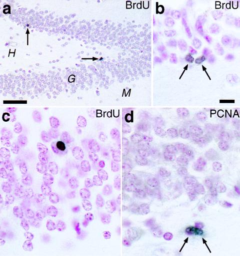
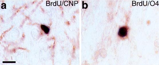
We present evidence for continuous generation of neurons, oligodendrocytes, and astrocytes in the hippocampal dentate gyrus of adult macaque monkeys, using immunohistochemical double labeling for bromodeoxyuridine and cell-type-specific markers. We estimate that the relative rate of neurogenesis is approximately 10 times less than that reported in the adult rodent dentate gyrus. Nevertheless, the generation of these three cell types in a discreet brain region suggests that a multipotent neural stem cell may be retained in the adult primate hippocampus. This demonstration of adult neurogenesis in nonhuman Old World primates-with their phylogenetic proximity to humans, long life spans, and elaborate cognitive abilities-establishes the macaque as an unexcelled animal model to experimentally investigate issues of neurogenesis in humans and offers new insights into its significance in the adult brain.


Figures

Figure 1
Figure 1
The presence of newly generated cells in the dentate gyrus of adult macaque monkeys, as revealed by independent immunohistochemical markers. (a and b) The hippocampal dentate gyrus 2 hr after the last of five daily injections of BrdU shows BrdU-labeled nuclei (arrows) in the subgranular zone of the granule cell layer (G). H, hilus; M, molecular layer. (b) A BrdU-labeled “doublet” in the SGZ, possibly the daughter cell progeny of a recent mitotic event. (c) A BrdU-labeled nucleus deep in the GCL, 27 days after a single BrdU injection. Note the rounded appearance, similar to that of neighboring granule neurons and oligodendrocytes. (d) An example of a PCNA-immunopositive doublet in the SGZ. [Bars = (a) 50 μm; (b–d) 10 μm.]
Figure 2
Figure 2
Newly generated cells in the adult macaque dentate gyrus express neuronal phenotypic markers 32 days after five BrdU injections, as detected by immunofluorescence double-label and confocal microscopy. (a–d) Neurons in the dentate gyrus express NeuN (red). The same cell in the GCL that is labeled with BrdU (arrow, green in b) also expresses NeuN (arrow, a). (c and d) An example of a BrdU-labeled nucleus (d, arrow, green) that did not emit a red fluorescence signal (c, arrow), demonstrating that the BrdU fluorescent signal did not “bleed” into the red channel; this might be a progenitor or new glial cell. (e and f) A TuJ1-positive cell in the SGZ (arrow, red) colabels with BrdU in its nucleus (f, arrow, green). Note the slender process (arrowheads) emanating from the cell body, resembling the trailing process of a newly generated migrating neuron. The BrdU in its nucleus confirms its recent generation. (g and h) Two cells in the SGZ expressing TuJ1 in the cytoplasm surrounding their nuclei (red), which are immunopositive for BrdU (h, green). Their close proximity suggests that these two cells might be newly generated “siblings.” The long thin process (arrowheads), consistent with migratory behavior, is clearly seen in one of the cells. (i and j) A bipolar cell in the SGZ coexpressing TuJ1 (green) and nuclear BrdU (j, orange). Although most double-labeled cells were oriented radially in the GCL, occasionally a cell was oriented parallel to the GCL. This example shows such a BrdU-labeled cell with an extended process on either side of the nucleus. (k) A TuJ1-positive cell (green, arrow) with a BrdU-positive nucleus (orange) has an immature migratory appearance. Note the thin trailing process (arrowheads) and a nearby BrdU-negative neuron, with a mature, apical process (arrow–cross). (l) A cell deep in the GCL colabels with TuJ1 (green) and BrdU (orange) with an apical process that is thick and tortuous, similar to the dynamic, exploratory leading process of a migrating neuron (its trailing process is out of the optical plane). Compare this with the straighter apical process of the more mature BrdU-immunonegative granule neuron in k (arrow–cross). [Bar (a–l) = 10 μm.]
Figure 3
Figure 3
Newly generated oligodendrocytes in the adult macaque dentate gyrus. These two examples show cells that are immunoperoxidase double-labeled; the black reaction product in the nucleus indicates BrdU, and the brown stain in the surrounding cytoplasm indicates the expression of oligodendrocyte-specific markers, CNP (a) or O4 (b). The cell in (a) is in the GCL and the cell in (b) is in the hilus of an adult monkey, 27 days after a single BrdU injection. (Bar = 10 μm.)

References

    1. Leblond C P. Natl Cancer Inst Monogr. 1964;14:119–150. - PubMed
    1. Rakic P. Science. 1985;227:1054–1056. - PubMed
    1. Altman J, Bayer S A. In: Restorative Neurology, Vol. 6, Neuronal Cell Death and Repair. Cuello A C, editor. Amsterdam: Elsevier; 1993. pp. 203–225.
    1. Altman J, Das G D. J Comp Neurol. 1965;124:319–336. - PubMed
    1. Kaplan M S, Hinds J W. Science. 1977;197:1092–1094. - PubMed

Publication types

Substances

LinkOut - more resources