. 1999 May 11;96(10):5774-9.
doi: 10.1073/pnas.96.10.5774.

Poly(ADP-ribose) polymerase activation mediates 1-methyl-4-phenyl-1, 2,3,6-tetrahydropyridine (MPTP)-induced parkinsonism

Affiliations

Poly(ADP-ribose) polymerase activation mediates 1-methyl-4-phenyl-1, 2,3,6-tetrahydropyridine (MPTP)-induced parkinsonism

A S Mandir et al. Proc Natl Acad Sci U S A. .

Abstract

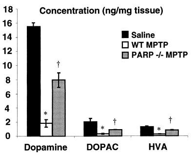
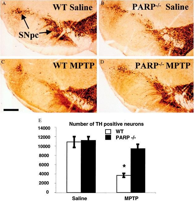
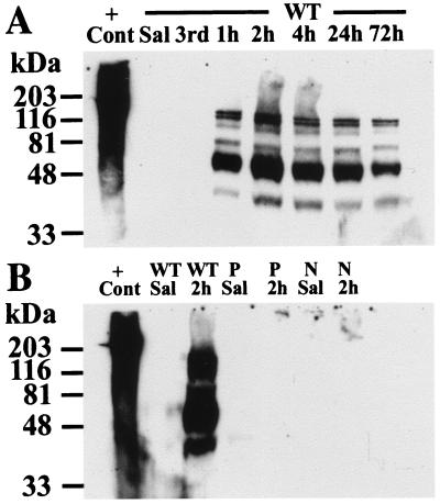
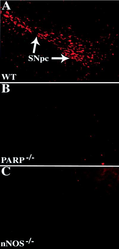
1-Methyl-4-phenyl-1,2,3,6-tetrahydropyridine (MPTP) is a neurotoxin that causes parkinsonism in humans and nonhuman animals, and its use has led to greater understanding of the pathogenesis of Parkinson's disease. However, its molecular targets have not been defined. We show that mice lacking the gene for poly(ADP-ribose) polymerase (PARP), which catalyzes the attachment of ADP ribose units from NAD to nuclear proteins after DNA damage, are dramatically spared from MPTP neurotoxicity. MPTP potently activates PARP exclusively in vulnerable dopamine containing neurons of the substantia nigra. MPTP elicits a novel pattern of poly(ADP-ribosyl)ation of nuclear proteins that completely depends on neuronally derived nitric oxide. Thus, NO, DNA damage, and PARP activation play a critical role in MPTP-induced parkinsonism and suggest that inhibitors of PARP may have protective benefit in the treatment of Parkinson's disease.


Figures

Figure 1
Figure 1
PARP−/− mice are resistant to the toxic effects of MPTP. HPLC with electrochemical detection of DA and metabolites, HVA and DOPAC, 1 week after MPTP administration. All animals are on a pure genetic background of 129 SvEv. No differences in striatal DA or metabolite content was observed in WT or PARP−/− animals after saline injection, and these values are graphically combined (n = 10). WT animals (n = 8) injected with MPTP (20 mg/kg ×4) have significant reductions in DA and metabolites compared with saline controls and PARP−/− (n = 5). Two-way ANOVA, P < 0.004 for DA, HVA, and DOPAC. Student-Newman-Keuls post hoc analysis, ∗, P < 0.05 WT MPTP vs. saline, , P < 0.05 WT MPTP vs. PARP−/− MPTP.
Figure 2
Figure 2
DA neurons from PARP−/− mice are resistant to MPTP neurotoxicity. TH immunostaining of representative midbrain sections 7 days after MPTP adminstration from (A) saline-injected WT, (B) saline-injected PARP−/−, (C) MPTP-injected WT, and (D) MPTP-injected PARP−/− mice. (E) A significant reduction of TH-immunopositive neurons is seen in the WT mice receiving MPTP (n = 5) compared with saline controls (n = 8) (∗, ANOVA with Fisher post hoc, P < 0.0001 WT MPTP vs. saline). No statistical difference is seen between saline controls and PARP−/− (n = 4) 1 week after MPTP administration (ANOVA). Counts of Nissl-stained neurons in midbrain yielded similar results (data not shown).
Figure 3
Figure 3
MPTP induces marked levels of poly(ADP-ribosyl)ation of nuclear proteins. Immunoblot analysis with mAb to poly(ADP-ribose). (A) No evidence of poly(ADP-ribosyl)ation is seen until after the fourth injection of MPTP. Polymer formation peaked at 2 hr after the fourth dose and is still detectable at 72 hr. (B) No polymer formation is seen in PARP−/− or nNOS−/− mice. No polymer formation is seen in the striata of WT animals after MPTP (not shown). P, PARP−/−; N, nNOS−/−; 3rd, time at the third injection of MPTP; h, hours after the fourth dose of MPTP; +Cont, sonicated Hela cells plus NAD as a positive control. This experiment has been replicated three times with similar results.
Figure 4
Figure 4
PARP is activated in DA neurons after MPTP intoxication. Immunohistochemical staining with an anti-poly(ADP-ribose) antibody (pseudocolored in red) in the ventral midbrain. (A) WT after MPTP delivery demonstrates intense and specific staining of DA neurons. (B) PARP−/− midbrains are devoid of immunostaining. (C) nNOS−/− mice lack poly(ADP-ribose) formation after MPTP. Poly(ADP-ribose) is not detectable in saline-injected animals (data not shown). These images were obtained from animals 4 hr after the fourth injection of MPTP. These experiments have been replicated three times with similar results.

References

    1. Hornykiewicz O. Clin Neurol Neurosurg. 1992;94:S9–S11. - PubMed
    1. Fahn S. Cecil’s Textbook of Medicine. Philadelphia: Saunders; 1988.
    1. Youdim M B, Riederer P. Sci Am. 1997;276:52–59. - PubMed
    1. Olanow C W. Trends Neurosci. 1993;16:439–444. - PubMed
    1. Forno L S, DeLanney L E, Irwin I, Langston J W. Adv Neurol. 1993;60:600–608. - PubMed

Publication types

MeSH terms

LinkOut - more resources