. 1999 May 11;96(10):5791-6.
doi: 10.1073/pnas.96.10.5791.

TRP2: a candidate transduction channel for mammalian pheromone sensory signaling

Affiliations

TRP2: a candidate transduction channel for mammalian pheromone sensory signaling

E R Liman et al. Proc Natl Acad Sci U S A. .

Abstract

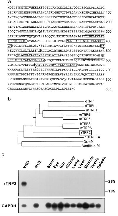
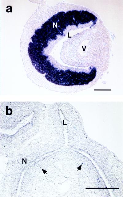
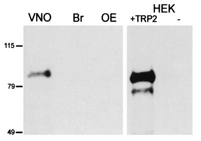
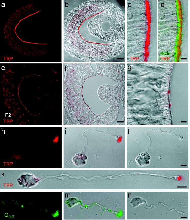
The vomeronasal organ (VNO) of terrestrial vertebrates plays a key role in the detection of pheromones, chemicals released by animals that elicit stereotyped sexual and aggressive behaviors among conspecifics. Sensory transduction in the VNO appears unrelated to that in the vertebrate olfactory and visual systems: the putative pheromone receptors of the VNO are evolutionarily independent from the odorant receptors and, in contrast to vertebrate visual and olfactory transduction, vomeronasal transduction is unlikely to be mediated by cyclic-nucleotide-gated channels. We hypothesized that sensory transduction in the VNO might instead involve an ion channel of the transient receptor potential (TRP) family, members of which mediate cyclic-nucleotide-independent sensory responses in Drosophila melanogaster and Caenorhabditis elegans and play unknown functions in mammals. We have isolated a cDNA (rTRP2) from rat VNO encoding a protein of 885 amino acids that is equally distant from vertebrate and invertebrate TRP channels (10-30% amino acid identity). rTRP2 mRNA is exclusively expressed in VNO neurons, and the protein is highly localized to VNO sensory microvilli, the proposed site of pheromone sensory transduction. The absence of Ca2+ stores in sensory microvilli suggests that, in contrast to a proposed mechanism of activation of mammalian TRP channels, but in accord with analysis of TRP function in Drosophila phototransduction, the gating of TRP2 is independent from the depletion of internal Ca2+ stores. Thus, TRP2 is likely to participate in vomeronasal sensory transduction, which may share additional similarities with light-induced signaling in the Drosophila eye.


Figures

Figure 1
Figure 1
rTRP2 is a VNO-specific ion channel of the TRP family. (a) Predicted amino acid sequence encoded by rTRP2 cDNA, showing the position of six putative transmembrane domains (solid outlines), and pore region (dotted underline). Stars indicate the positions of two mutations introducing stop codons within the ORF of the corresponding human sequence. (b) Phylogenetic tree of members of the TRP family in Drosophila (dTRP and dTRPL), mouse (mTRP1, -4, -5, and -6), human (hTRP3 and vanilloid R1), and C. elegans (OSM9 and KO1A11.4). Full-length protein sequences were aligned in CLUSTAL_X (42), and phylogeny was calculated by the neighbor-joining method, after removal of gapped regions, with 1,000 bootstrap trials. GenBank accession numbers are P19334 (dTRP), M88185 (dTRPL), U73625 (mTRP1), U50922 (mTRP4), AF029983 (mTRPS), N13758 (hTRP3), U49069 (mTRP6), Z66514 (K01A11.4), AF0115639 (OSM9), and AF029310 (VR1). (c) Northern blot analysis shows that rTRP2 transcripts are confined to the VNO.
Figure 2
Figure 2
rTRP2 is strongly expressed in adult rat VNO neurons. (a) In VNO cross sections hybridized to digoxigenin probe for rTRP2, the VNO neuroepithelium (N) appears strongly and specifically labeled. L, VNO lumen; V, VNO vein. (b) In the MOE, some sparse cells are faintly labeled (arrows) that are mostly located in the basal half of the neuroepithelium. N, neuroepithelium; L, lumen.
Figure 3
Figure 3
Detection of rTRP2 protein in immunoblots. Western blots were probed with an antibody raised against the carboxyl terminus of rTRP2. Lanes contain total protein (20 μg per lane) from VNO, brain (Br), and olfactory epithelium (OE), and from HEK cells transfected with TRP2 or untransfected (−).
Figure 4
Figure 4
Localization of rTRP2 to the sensory microvilli of vomeronasal neurons. (a and b) Adult VNO labeled with affinity-purified anti-rTRP2 antibody (red) and shown as the fluorescence signal (a) or a digital addition of the fluorescence and bright-field images (b). rTRP2 is located at the luminal surface of the sensory epithelium. (c and d) Higher magnification of the VNO sensory epithelium labeled with the anti-rTRP2 antibody (red) and Bodipy phalloidin to label actin (green). Label is shown as the red fluorescence channel combined digitally with the differential interference contrast (DIC) image (c), or both red and green channels combined with DIC. TRP2 is located mostly distal to the actin-rich terminal web. (e–g) Postnatal-day-2 VNO, labeled with anti-rTRP2 and shown as in a–c. Luminal labeling is present but more punctate than in adult. (hj) Localization of rTRP2 in microvilli, in a dissociated rat VNO neuron. Images are fluorescence (h), combined fluorescence and DIC (i), or DIC alone (j). Label is restricted to the microvilli and not apparent in the dendritic knob. (k) Another VNO neuron labeled as in i. (l–n) Localization of Gi2–3 immunoreactivity in the microvilli of a dissociated VNO neuron. (Scale bars: a and b, 100 μm; e and f, 50 μm; c, d, g, and h–n, 5 μm.)

Similar articles

Cited by

References

    1. Halpern M. Annu Rev Neurosci. 1987;10:325–362. - PubMed
    1. Schild D, Restrepo D. Physiol Rev. 1998;78:429–466. - PubMed
    1. Buck L, Axel R. Cell. 1991;65:175–187. - PubMed
    1. Zhao H, Ivic L, Otaki J M, Hashimoto M, Mikoshiba K, Firestein S. Science. 1998;279:237–242. - PubMed
    1. Bradley J, Li J, Davidson N, Lester H, Zinn K. Proc Natl Acad Sci USA. 1994;91:8890–8894. - PMC - PubMed

Publication types

MeSH terms

Associated data